
Fig. S2
- ID
- ZDB-FIG-070810-10
- Publication
- Zhang et al., 2007 - SCL-GFP transgenic zebrafish: In vivo imaging of blood and endothelial development and identification of the initial site of definitive hematopoiesis
- Other Figures
- All Figure Page
- Back to All Figure Page
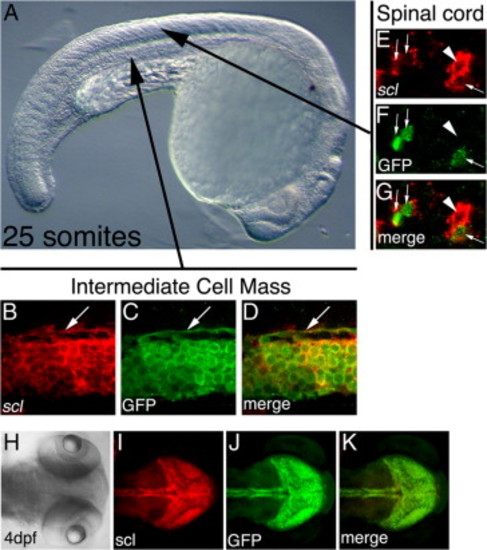
Colocalization of expression of endogenous scl message and GFP protein in scl-PAC-GFP transgenic embryos. scl message was detected by whole-mount in situ hybridization using the red fluorescent substrate, Fast Red (Sigma). Subsequently, GFP protein was detected using affinity-purified rabbit polyclonal antiserum to GFP (Torrey Pines Biolabs), followed by a goat anti-rabbit F(ab2)2 secondary antiserum labeled with AlexaFluor488 (Molecular Probes). Whole-mount in situ signal (red fluorescence) and immunofluorescence (green fluorescence) were imaged using a Nikon C1 confocal microscope. In general, expression of the GFP transgene coincides with the expression of the endogenous scl message. However, in tissues where scl expression has newly initiated, detection of the GFP protein is, as would be expected, delayed relative to detection of the scl message. (A) Lateral view of a 25-somite zebrafish embryo. Arrows indicate the regions imaged in panels B to G. (B?D) Intermediate cell mass, sagittal confocal slice. (B) Endogenous scl message. (C) GFP protein. (D) Overlay of panels B and C. GFP is co-expressed with the endogenous scl message in the mass of primitive erythroblasts as well as in the endothelium of the forming dorsal aorta (white arrow). (E?F) Ventral part of spinal cord, parasagittal confocal slice. (E) Endogenous scl message. (F) GFP protein. (G) Overlay of panels E and F. Expression of endogenous scl has recently initiated in interneurons in the ventral spinal cord. GFP protein can be detected in most (white arrows) but not all (white arrowhead) of these cells. (H?K) Dorsal view of head of 4 dpf embryo. Fluorescence images are Z-projections of confocal stacks. (H) Brightfield. (I) Endogenous scl message. (J) GFP protein. (K) Overlay of panels I and J. GFP protein is co-expressed with endogenous scl message in ventral hindbrain, optic tectum and diencephalon. |
| Gene: | |
|---|---|
| Fish: | |
| Anatomical Terms: | |
| Stage Range: | Prim-5 to Day 4 |
Reprinted from Developmental Biology, 307(2), Zhang, X.Y., and Rodaway, A.R., SCL-GFP transgenic zebrafish: In vivo imaging of blood and endothelial development and identification of the initial site of definitive hematopoiesis, 179-194, Copyright (2007) with permission from Elsevier. Full text @ Dev. Biol.