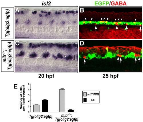
Loss of Notch signaling causes formation of excess PMNs and a deficit of KA' interneurons. (A-D) Lateral views of the spinal cord at the 6- to 12-somite region of zebrafish embryos, with anterior to left and dorsal at the top. (A) A subset of PMNs (CaP and VaP) and Rohon-Beard neurons of Tg(olig2:egfp) embryos expressed isl2 in the ventral spinal cord (black bracket) and dorsal spinal cord (white bracket) at 20 hpf, respectively. (B) In Tg(olig2:egfp) embryos, KA' interneurons (arrowheads) were detected by anti-GABA antibody (red) within EGFP+ cells at 25 hpf. EGFP- GABA+ cells (arrows) in ventral spinal cord are KA'' interneurons (Park et al., 2004). (C) In mib-/-;Tg(olig2:egfp) embryos, isl2+ PMNs formed as clusters in each hemisegment in the ventral spinal cord (black bracket). (D) mib-/-;Tg(olig2:egfp) embryos did not produce EGFP+ GABA+ KA' interneurons, although EGFP- GABA+ KA'' interneurons were still present (arrows). (E) Mean of the number of PMNs (gray bar) and KA' interneurons (black bar) per hemisegment in Tg(olig2:egfp) and mib-/-;Tg(olig2:egfp) embryos (n=7). Error bars represent s.e.m. Scale bar: 50 µm.
|
