
Fig. 5
- ID
- ZDB-FIG-070613-105
- Publication
- Georgijevic et al., 2007 - Spatiotemporal expression of smooth muscle markers in developing zebrafish gut
- Other Figures
- All Figure Page
- Back to All Figure Page
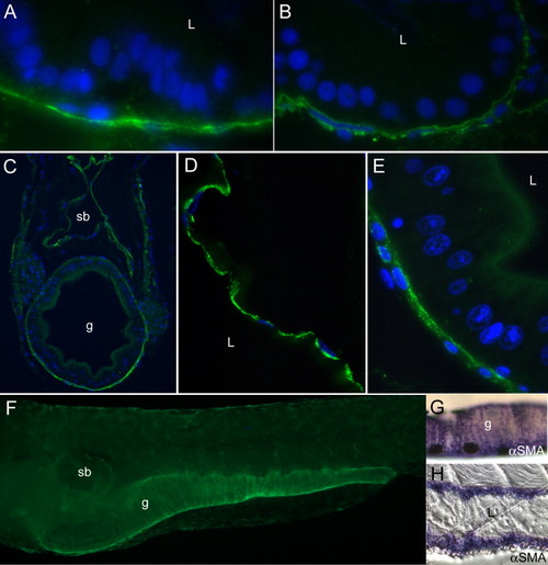
Smooth muscle marker expression in the juvenile gut and swim bladder. A,B: Tropomyosin is expressed in a circular smooth muscle layer at 4 days postfertilization (dpf; A), and in longitudinal and circular layers at 6 dpf (B). C-F: By15 dpf, tropomyosin staining is strong in a layer surrounding both the gut and swim bladder (C). D,E: A higher power image (D) reveals that the swim bladder has a thin epithelial lining and single layer of smooth muscle cells surrounding it, while the gut (E) at similar magnification has developed a columnar epithelium, and a thicker smooth muscle wall. F: In a whole-mount larva, tropomyosin staining is seen around the swim bladder and gut. G: Whole-mount staining of a 20 dpf larva with α-smooth muscle actin (αSMA) reveals a similar pattern to tropomyosin. H: Longitudinal sections of a 22 dpf larva show extensive smooth muscle layers expressing αSMA. L, lumen; sb, swim bladder; g, gut. |