
Fig. 2
- ID
- ZDB-FIG-070309-13
- Publication
- Sassa et al., 2007 - Visualization of two distinct classes of neurons by gad2 and zic1 promoter/enhancer elements in the dorsal hindbrain of developing zebrafish reveals neuronal connectivity related to the auditory and lateral line systems
- Other Figures
- All Figure Page
- Back to All Figure Page
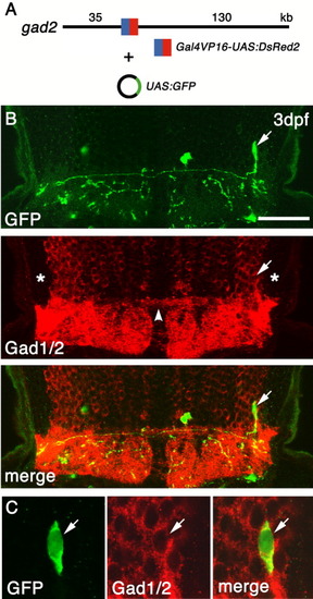
Visualization of a single Gad1/2(+) axon projecting to the contralateral hindbrain. A: Structure of the gad2 bacterial artificial chromosome (BAC) clone with Gal4VP16/UAS:DsRed2 inserted into the 5′-untranslater region (5′-UTR) of gad2. B: A coronal section of the hindbrain of an embryo injected with the modified gad2 BAC DNA and UAS:GFP plasmid DNA. Dorsal is to the top. A single axon of a green fluorescent protein-positive (GFP(+)) neuron (arrow) projects to the contralateral hindbrain. Asterisks indicate the regions likely occupied by the zn-5(+)Lhx2/9(+) neurons. Note the absence of Gad1/2 signal in this region. Note also the Gad1/2(+) commissural fascicles connecting the left and right lateral Gad1/2(+) neurons (arrowhead). C: The same single GFP(+) neuron as in B, at higher magnification, demonstrating that this neuron is also positive for Gad1/2. UAS, upstream activating sequences. Scale bars = 75 μm in B, 15 μm in C. |