|
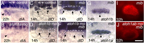
Interactions between atoh1 and the Delta-Notch pathway. (A,B) Expression of dlA at 22 hpf in a control embryo (A) and atoh1a morphant (B). (C-F) Expression of dlD at 14 hpf in a control embryo (C), atoh1b morphant (D), mib mutant (E) and mib mutant-atoh1b morphant (F). (G,H) mib mutants show expanded otic domains of atoh1b (G) and atoh1a (H) at 14 hpf. (I,J) Pax2 antibody staining at 32 hpf reveals supernumerary hair cells in a mib mutant (I) but no hair cells in a mib mutant co-injected with atoh1a MO and atoh1b MO (J). Arrowheads and arrows indicate otic regions. All images are dorsolateral views with anterior to the left. Scale bars: 30 μm in A,E,I-P; 15 μm in B-D,F-H.
|