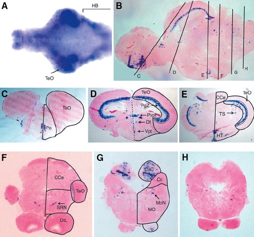
Expression of FoxP2 in the zebrafish brain. (A) Dorsal view of in situ hybridization of FoxP2 probe to the isolated brain from a 7 day old zebrafish. The isolated brain was opened along its dorsal axis and flattened. Anterior is to the left. (B) Sagittal section of a brain from a 3 months old zebrafish hybridized with a FoxP2 probe. Vertical lines indicate the positions of cross sections in images (C - H). Cross sections, hybridized with a FoxP2 probe, through the (C) telencephalon, (D) optic tectum, (E) optic tectum, cerebellum and hypothalamus, (F) cerebellum, (G) caudal lobe of the cerebellum and the medulla oblongata. Arrow in (F) indicates the expression in the superior reticular formation. Arrow in (G) indicates expression in the medial octavolateralis nucleus. (H) A section caudal to (G) shows no expression of FoxP2. Abbreviations: Cc, cerebellar crest; CaC, caudal lobe of cerebellum; CCe, corpus cerebelli; DIL, diffuse nucleus of the inferior hypothalamic lobe; Dt, dorsal thalamus; HB, hindbrain; HT, hypothalamus; MO, medulla oblongata; MoN, medial octavolateralis nucleus; PgZ, periventicular gray zone of the optic tectum; Po, preoptic area; Pvpt, periventricular pretectum; SRN, superior reticular nucleus; TeO, optic tectum; TS, torus semicircularis; Vpt, ventral posterior tuberculum.
|
