
Fig. 3
- ID
- ZDB-FIG-060209-4
- Publication
- Mao et al., 2006 - Developmentally regulated gene expression of the small heat shock protein Hsp27 in zebrafish embryos
- Other Figures
- All Figure Page
- Back to All Figure Page
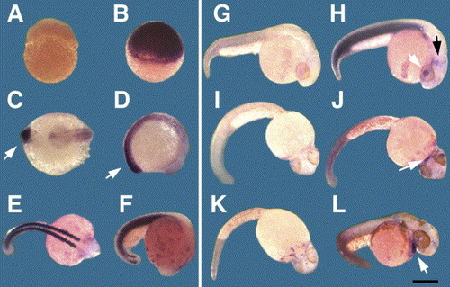
Detection of hsp27 expression in whole embryos during normal development. Antisense, Hsp27 mRNA specific probe (A?F,H,J,L) and a sense control probe (G,I,K) were used for hybridization. Embryos are shown at (A) high blastula, (B) 30% epiboly, (C,D):19, (E,F): 24, (G,H):36, (I,J):48, (K,L):78 hpf. Images were obtained after overnight colorimetric development, except a 1hr development was used for embryos collected at the tail bud stage (C,D). Arrows indicate the posterior tip of the embryo (C,D), midbrain/hindbrain (black arrow, H), lens (white arrow, H) and heart (J,L). Images are oriented as described in Experimental Procedures, except that the dorsal surface of the embryo is shown in E and the ventral surface is shown in C,I,J,K. Bar=300 μm. |
| Gene: | |
|---|---|
| Fish: | |
| Anatomical Terms: | |
| Stage Range: | 30%-epiboly to Protruding-mouth |
Reprinted from Gene expression patterns : GEP, 6(2), Mao, L., and Shelden, E.A., Developmentally regulated gene expression of the small heat shock protein Hsp27 in zebrafish embryos, 127-133, Copyright (2006) with permission from Elsevier. Full text @ Gene Expr. Patterns