
Fig. 2
- ID
- ZDB-FIG-060203-1
- Publication
- Murakami et al., 2006 - Zebrafish protocadherin 10 is involved in paraxial mesoderm development and somitogenesis
- Other Figures
- All Figure Page
- Back to All Figure Page
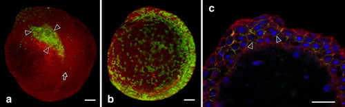
Ectopic expression of tagged Pcdh10 in zebrafish embryos. Single blastomeres of eight-cell embryos were injected with an RNA of GFP-tagged Pcdh10 construct, Pcdh10G. a: Confocal microscopy of 11 hours postfertilization (hpf) embryos expressing Pcdh10G revealed that fluorescent cells remained coherent during gastrulation to form patches (arrowheads) on the embryo. In this particular embryo, the left segmental plate was split from the notochord (arrow) at such a patch of green cells. GFP image (green) was superimposed on a phalloidin image (red). Left-side oblique view with rostral to the top. b: Confocal image of a 11 hpf embryo injected with GFP control RNA showed green cells dispersed over the embryo. Left-side view with rostral to the top. c: Detailed confocal scan of a 9 hpf embryo showing Pcdh10G localized to the cell peripheries (green, arrowheads). Red, phalloidin; blue, 4,6-diamidino-2-phenylindole (DAPI). The animal pole is to the top, dorsal to the right. a and b, Z projections of serial optical sections. c, a single section. Scale bars = 50 μ m. |