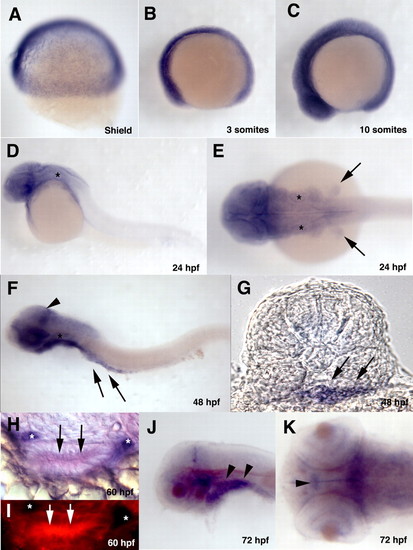
Developmental expression pattern of trap100. (A-F) Whole-mount in situ hybridized embryos hybridized with a trap100 antisense probe at the indicated developmental stages. (A-D,F) Lateral views; (E) Dorsal view of 24 hpf embryo. Asterisks in D-F indicates the otic vesicle. Arrows in E indicate fin buds. Arrowhead in F indicates increased expression of trap100 in the posterior mesencephalon. Arrows in F indicate intestinal mesendodermal expression of trap100. (G) A transverse section taken through 48 hpf embryo at the level of somite 4 showing expression through the intestinal mesendoderm. Arrows indicate intestinal mesendodermal expression of trap100. (H) A transverse section taken trough the gut tube at 60 hpf after double in situ hybridization with a trap100 fluorescein antisense probe (red) and a phox2b digoxigenin antisense probe (purple) showing intestinal epithelia expression of trap100. (I) Same section as in H showing trap100 expression using fluorescence in the intestinal epithelia cells. Arrows and asterisks in H,I indicate intestinal mesendodermal expression of trap100and phox2b-positive ENS precursors, respectively. (J,K) Whole-mount in situ hybridized embryos hybridized with a trap100 antisense probe at 72 hpf. (J) Lateral view; (K) dorsal view. Arrowheads in J indicate pharyngeal arch mesendodermal expression. Arrowhead in K indicates ventral diencephalon cells expressing neurons expressing trap100. In all whole mounts (A-F,J,K), anterior is towards the left; (F,K,J) yolk has been removed.
|
