
FIGURE
Fig. S1
- ID
- ZDB-FIG-050729-10
- Publication
- Xiao et al., 2005 - A GFP-based genetic screen reveals mutations that disrupt the architecture of the zebrafish retinotectal projection
- Other Figures
- All Figure Page
- Back to All Figure Page
Fig. S1
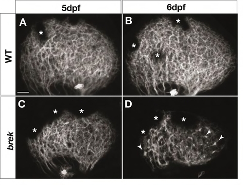
Retinal axons degenerate in breaking up (brek) mutants. Z-projections of confocal image stacks showing lateral views of the tectum in 5-6 dpf live Brn3c:mGFP larvae. At 5 dpf, retinal axons cover the tectum normally in brek mutants (C), similar to wild type (A). At 6 dpf, retinal axons become punctate in the brek tectum and coverage is more sparse (D) than in wild type (B). Arrowheads indicate puncta. Asterisks indicate melanophores in the skin. Scale bars: 20 mm. |
Expression Data
Expression Detail
Antibody Labeling
Phenotype Data
Phenotype Detail
Acknowledgments
This image is the copyrighted work of the attributed author or publisher, and
ZFIN has permission only to display this image to its users.
Additional permissions should be obtained from the applicable author or publisher of the image.
Full text @ Development