Fig. 7
- ID
- ZDB-FIG-050715-11
- Publication
- Rhinn et al., 2005 - Positioning of the midbrain-hindbrain boundary organizer through global posteriorization of the neuroectoderm mediated by Wnt8 signaling
- Other Figures
- All Figure Page
- Back to All Figure Page
|
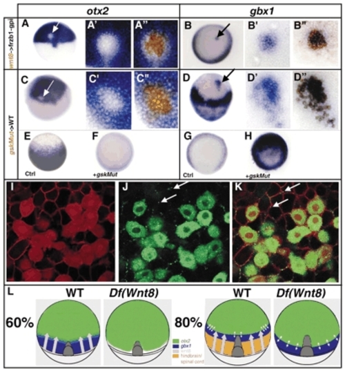
Wnt8 represses otx2 and induces gbx1 in a non-cell-autonomous manner. (A-B″) Embryos containing cells derived from injected embryos with a lineage tracer (brown) and wnt8 RNA (400 pg) into the animal pole of a host embryo injected with Fzb1-gpi RNA (200 pg). (C-H) A dominant-negative gsk-3 can activate the Wnt pathway in a cell-autonomous manner. Embryos containing cells derived from injected embryos with a lineage tracer (brown) and Xgsk-3K→R (gskMut) RNA (400 pg). (A-A″,C-C″) Embryos stained for otx2 expression. Close-up of the transplanted cells (A′-C′) before biotin staining and (A″-C″) after biotin staining. otx2 repression occurs in the transplanted cells only and not in the surrounding host tissue. (B-B″,D-D″) Embryos stained for gbx1 expression. Close-up of the transplanted cells (B′-D′) before biotin staining and (B″-D″) after biotin staining. gbx1 is induced in the transplanted cells only. (E-H) Animal pole views. (E) Control embryos at 60% stained for otx2. (F) gskMut-injected embryos do not express otx2. (G) Control embryos at 60% stained for gbx1. (H) gskMut-injected embryos express gbx1 ectopically mimicking ectopic wnt expression. (I-K) Embryos containing cells derived from injected embryos with a lineage tracer (red in the whole cell in I) and wnt8-gfp mRNA (green in J) (800 pg). The host embryos injected with palmitoylated mRFP mRNA (100 pg), which labels the cell membrane in red (I). (K) Overlay of (I) and (J). White arrows indicate the Wnt8 protein that has been secreted from the donor cells. (L) Wnt8 graded expression inhibits otx2 and induces gbx1 expression in the most posterior region of the embryos at 60% of epiboly. At this stage, the otx2 and gbx1 expression domains overlap slightly. Loss of Wnt8 leads to the loss of gbx1 expression and to a posterior shift of the otx2 expression domain. At 80% of epiboly, the otx2 and gbx1 expression domains are sharp and complementary, probably owing to mutual repressive interactions. In absence of Wnt8, the gbx1 expression domain is established with a posterior shift. Its expression is complementary to the otx2 expression domain. |

