
Fig. 1
- ID
- ZDB-FIG-050621-15
- Publication
- Zhu et al., 2005 - Regulation of the lmo2 promoter during hematopoietic and vascular development in zebrafish
- Other Figures
- All Figure Page
- Back to All Figure Page
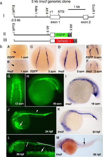
EGFP expression under the control of a 2.5-kb lmo2 promoter recapitulates endogenous lmo2 expression. (a) Constructs used to generate the lmo2 transgenic lines are shown. Construct I is a 5-kb genomic fragment derived from a PAC clone that contains exons 1 and 2 of lmo2. Constructs II and III are 2.5 kb of the 5-kb fragment ligated to the reporter genes EGFP-1 and DsRed2-1. (b) EGFP and lmo2 transcripts first appear in two lateral stripes of ventral mesoderm (black arrowheads) by the 1-somite stage as shown in A and B. Transcripts can been detected at the 5-somite stage (C, D). Anterior vascular precursors express EGFP and lmo2 at 5 somites (E, F). At 12 somites, the posterior mesoderm stripes migrate to form the ICM (G). By the 18-somite stage, most of the ICM has formed from the convergent morphogenesis of the stripes (H, I). At 24 hpf, circulating blood (white arrow) and most of the vasculature, including the cardinal vein, dorsal aorta, and branching intersomitic vessels (white arrowhead), are fluorescent (J), which replicate that of endogenous lmo2 expression in K. In line LGb, EGFP is downregulated over the next 24 h, but vascular expression persists (L); the aorta and vein are fluorescently labeled during the formation of the presumptive AGM at 36 hpf (inset and white arrow). Endogenous lmo2 is expressed at low levels in the trunk vessels at 36 hpf (black arrow) (M). |
| Gene: | |
|---|---|
| Fish: | |
| Anatomical Terms: | |
| Stage Range: | 1-4 somites to Prim-25 |
Reprinted from Developmental Biology, 281(2), Zhu, H., Traver, D., Davidson, A.J., Dibiase, A., Thisse, C., Thisse, B., Nimer, S., and Zon, L.I., Regulation of the lmo2 promoter during hematopoietic and vascular development in zebrafish, 256-269, Copyright (2005) with permission from Elsevier. Full text @ Dev. Biol.