This material is from the 4th edition of The Zebrafish Book. The 5th edition is available in print and within the ZFIN Protocol Wiki.
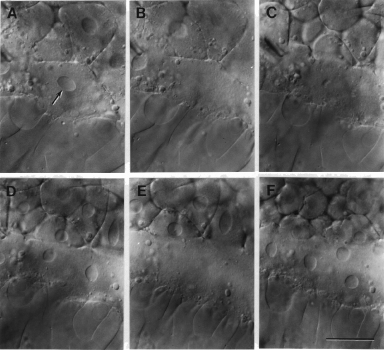
Fig 10. Formation of the yolk syncytial layer (YSL), viewed with Nomarski optics. A: A marginal blastomere during the tenth interphase (512-cell stage; the arrow indicates its nucleus). B: The nucleus disappears as the cell enters the tenth mitosis, a special one because there is no cytokinesis. The daughter nuclei then reappear in a common cytoplasm during interphase 11. During very early interphase the nuclei are globular (C), and later, just before entering mitosis again they are large and football-shaped (D). The YSL nuclei continue to disappear during mitoses (E; mitosis 11; note that interphase nuclei are present just in the blastoderm cells and not the YSL, meaning that the blastoderm and YSL are no longer in synchrony,) and reappear during interphases (F; interphase 12) for several mitotic cycles, always within a common syncytium. From Kimmel and Law (1985b). Scale bar: 50 µm.


Figure 10