- Title
-
Spadetail-dependent cell compaction of the dorsal zebrafish blastula
- Authors
- Warga, R.M. and Nüsslein-Volhard, C.
- Source
- Full text @ Dev. Biol.
|
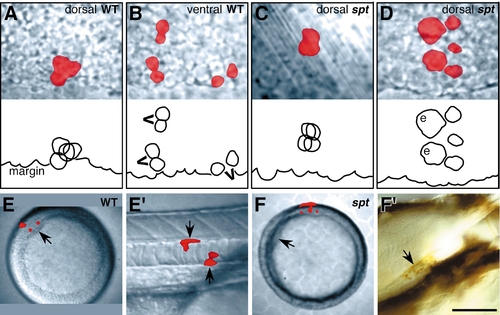
Cell clustering and dorsoventral position of prospective axial mesendoderm is altered in spadetail mutants. (A–D) Labeled clones at 40% epiboly. The upper panel is the record, and the lower panel is a tracing of the individual deep cells, typically by this time there were 4 deep cells descended from the mid-blastula marginal cell (3.7 cells ± 0.22, wild type; 3.9 cells ± 0.39, spadetail mutant); blastoderm margin is indicated; enveloping layer cells (e); recent cell divisions (v’s). Dorsal clones in mutant embryos disperse significantly more than in normal embryos (χ2 = 21.1, df = 4; P < 0.01). (E and F) Labeled clones contributing to notochord in the trunk. (E and F) Animal pole views at shield stage; dorsal side (arrow). (E′ and F′) Side views. (E′) 1 day wildtype embryo. The differentiated clone (arrows), located midtrunk, is notochord sheath, a derivative of the notochord (Melby et al., 1996). (F′) 4 day spadetail-mutant embryo, after whole-mount staining for the fixable tracer. The differentiated clone, now stained brown (arrow), is notochord sheath located in the anterior trunk. Scale bar: 50 μm (A–D), 100 μm (E′ and F′), 250 μm (E and F). |
|
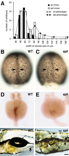
There are more axial and less paraxial cells in spadetail mutants. (A) The bimodal distribution in the width of the dorsal axis for embryos derived from a cross of spadetail heterozygotes (open bars), and the bell curve for embryos derived from a cross of wildtype homozygotes (solid bars). Overlap is apparent between the larger peak in the spadetail cross and the wildtype peak, suggesting that embryos within this region of the graph are phenotypically wildtype. The smaller peak in the spadetail cross would represent mutant embryos. Black lines with open or closed circles are the inferred distribution based on the expected mutant frequency. Axis were measured on a dissection scope using an ocular micrometer. (B and C) Dorsal axis (arrows) visualized using anti-Fkd2 immunostaining at 80% epiboly. (B) Representative wildtype embryo. (C) Presumed mutant embryo. (D and E) The embryonic gut in 1-day embryos visualized using fkd2 RNA expression (Odenthal and Nüsslein-Volhard, 1998). (F and G) The differentiated gut in 5 day embryos, the stomach (white arrow), intestine (black arrow), and swimbladder (small black arrow), all derived from the paraxial region (Warga and Nüsslein-Volhard, submitted) are present, but diminutive in the mutant. Note that muscle is now apparent, but less abundant in the mutant trunk as revealed by birefringence (open arrow). Scale bar: 60 μm (B and C), 100 μm (D–G). |
|
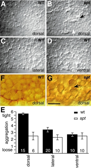
The behavior of blastula marginal cells is changed in spadetail mutants. (A–D) Marginal cells at 40% epiboly, after progression of cell compaction. (E) Assessment of appearance of marginal deep cells just below the surface of the enveloping layer between 30 and 40% epiboly, respective of position in wildtype (solid bar) and spadetail mutants (open bar); lines represent standard error. For reference we ranked A as 6, tightly associated cells, and B and C as 3, tightly and loosely associated cells, and D as 2, loosely associated cells. Cell compaction was statistically compared between wildtype dorsal and lateral cells (Wilcoxon rank sum = 150, n = 15, m = 20) and between wild-type dorsal and mutant dorsal cells (Wilcoxon rank sum = 120, n = 15, m = 6). (F and G) Cell membranes visualized using anti-β-catenin immunostaining at 30% epiboly. Scale bar: 50 μm (A–D), 25 μm (F and G). |
Reprinted from Developmental Biology, 203, Warga, R.M. and Nüsslein-Volhard, C., Spadetail-dependent cell compaction of the dorsal zebrafish blastula, 116-121, Copyright (1998) with permission from Elsevier. Full text @ Dev. Biol.


