
- Title
-
Membrane progesterone receptor γ (paqr5b) is essential for the formation of neurons in the zebrafish olfactory rosette
- Authors
- Mustary, U.H., Maeno, A., Rahaman, M.M., Ali, M.H., Tokumoto, T.
- Source
- Full text @ Sci. Rep.
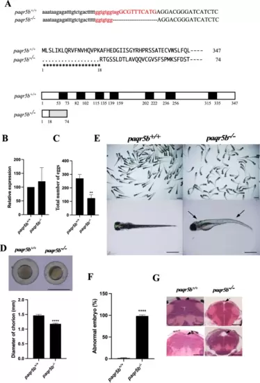
Fertility and embryonic development of the established paqr5b?/? line. (A) DNA sequence and predicted protein structure of an established paqr5b gene-edited strain. (Uppest panel) DNA sequences around the target site (in red) for CRISPR/Cas9 digestion in wild-type (paqr5b+/+) and paqr5b?/? mutants are indicated. A 13-nucleotide deletion was induced in the target site in the selected mutant. (Middle panel) The predicted protein sequences of the paqr5b+/+ and paqr5b?/? mutants are indicated. It was expected that a peptide of 74 amino acids in length with an N-terminal 18 amino acid sequence produced in the paqr5b?/? mutant was the same as that produced in wild-type Paqr5b zebrafish. Thus, only an N-terminal short fragment without a transmembrane region is present in the Paqr5b protein produced in the paqr5b?/? mutant. (Lowest panel) Diagrams of the predicted protein structures of the paqr5b+/+ and paqr5b?/? mutant strains are shown. The predicted transmembrane regions and the altered amino acid regions in the paqr5b?/? mutant are indicated by black boxes and diagonal boxes, respectively. (B) The relative expression levels of paqr5b in the olfactory rosette (OR) of the wild-type and the paqr5b mutants were compared. mRNA abundance was measured in triplicate for each sample from 10 fish, and all data from three preparations were normalized by the number of elongation factor 1? (EF1?) transcripts in each sample. The data indicated the expression level of the wild type set as 100. (C) Fecundity of paqr5b+/+ and paqr5b?/? zebrafish. The total number of eggs crossing each line was counted (n = 4, ** P ? 0.001). (D) Representative photograph of developing embryos with different sizes of chorions. The scale bar is 1 mm. Comparison of choroidal diameter between paqr5b+/+ and paqr5b?/? embryos (n = 4, **** P ? 0.00001). (E) Representative photograph of embryos at 5 dpf. Typical head abnormalities and spine curvatures in the paqr5b?/? embryos are indicated by arrows. The scale bars in the upper panels are 1 mm. The scale bars in the lower panels are 500 ?m. (F) Comparison of the percentage of abnormal embryos between the paqr5b+/+ and paqr5b?/? embryos (n = 4, **** P ? 0.00001). (G) Comparison of brain transverse sections of paqr5b+/+ and paqr5b?/? embryos. The ventricle of the embryo brain is indicated by arrows. The scale bars are 20 ?m. |
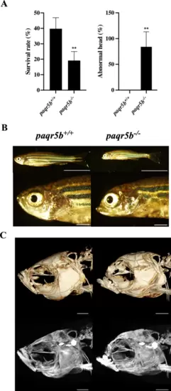
Abnormality of head structure in the paqr5b?/? mutant. (A) Survival rates of adult paqr5b+/+ and paqr5b?/? mutant fish (F6 generation). The number of surviving adult paqr5b+/+ and paqr5b-/- fish (left panel) and the percentage of head abnormalities (right panel) in each of the three batches were compared. Each value represents the mean of the data, and the vertical lines indicate the standard deviations. Asterisks represent significant differences between paqr5b+/+ and paqr5b?/? fish (* P ? 0.01). (B) Representative morphologies of the head region of adult paqr5b+/+ and paqr5b?/? fish are shown. The scale bars in the upper panels are 1 cm. The scale bars in the lower panels are 1 mm. (C) Micro-CT scan of the head skeleton of paqr5b+/+ and paqr5b?/?. The upper panels are volume-rendered images, and the lower panels are maximum-intensity projection images. Scale bars are 1 mm. PHENOTYPE:
|

Abnormal morphology of the brain in paqr5b?/?. (A) Comparison of the morphology of dissected brains between paqr5b+/+ and paqr5b?/? fish. The scale bars in the upper panels are 200 ?m. The scale bars in the lower panels are 100 ?m. Sagittal sections (B) and transverse sections (C) of the heads of paqr5b+/+ and paqr5b?/? are shown. The olfactory bulb (OB), telencephalon (Tel), tectum opticum (TeO) and corpus cerebeli (CCe) are indicated. Scale bars are 200 ?m in sagittal sections. The scale bars are 50 ?m in transverse sections. PHENOTYPE:
|
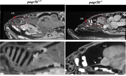
Observation of soft tissues inside the head region by micro-CT. Sagittal micro-CT images of the head region of adult paqr5b+/+ and paqr5b?/? are shown. The olfactory mucosa (OM), olfactory bulb (OB), telencephalon (Tel), tectum opticum (TeO) and corpus cerebeli (CCe) are indicated. Connections between the OM and OB are indicated by arrowheads. The scale bars in the upper panels are 500 ?m. The scale bars in the lower panels are 100 ?m. PHENOTYPE:
|

Expression of Paqr5b in olfactory mucosa (OM). (A) Expression analysis of Paqr5b in the OM. Proteins in the OM of paqr5b+/+ and paqr5b?/? zebrafish were detected by Coomassie Brilliant Blue R staining (CBBR). Paqr5b in the OM was detected with an anti-zebrafish Paqr5b antibody (a-Paqr5b). The arrowhead indicates a protein band corresponding to Paqr5b. (B) Immunohistochemical observation of Paqr5b in OM. Immunohistochemical staining results for Paqr5b in paqr5b+/+ and paqr5b?/? OMs. Sagittal sections of the OM of paqr5b+/+ and paqr5b?/? zebrafish were stained with an anti-zebrafish Paqr5b antibody (a-Paqr5b) and DAPI. Images of DAPI signals observed through a DAPI filter (DAPI) and images of immunostaining signals observed through the FITC filter. Merged images (Merge) of differential interference contrast (DIC) images and images of DAPI and anti-Paqr5b (a-Paqr5b) staining are shown. Enlarged images of DAPI and anti-Paqr5b-stained sections are indicated by red squares (lower panels). Neuronal cells (Crypt, Microvillus and Ciliated) that reacted with anti-Paqr5b in paqr5b+/+ are indicated by arrows. These OSNs are absent in paqr5b?/?. Nonspecific staining with the anti-Paqr5b antibody is indicated by a white arrowhead. The scale bars indicate 200 ?m in the upper panels and 50 ?m in the enlarged images. (C) Sagittal sections of paqr5b+/+ and paqr5b?/? were stained with hematoxylin, eosin and Alcian blue (H-E-A). The stained glycan layers are indicated by arrows. Scale bars are 200 ?m in the upper panels and 50 ?m in the lower panels. |
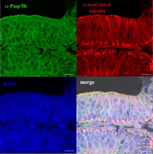
Paqr5b is expressed in neurons in the OR. Immunofluorescence staining of sections of OR from paqr5b+/+. Sections were double stained with anti-rabbit Paqr5b polyclonal antibodies (?-Paqr5b), anti-acetylated tubulin monoclonal antibody (?-acetylated tubulin) and DAPI. Merged images of both antibodies and DAPI are also shown (merge). Scale bars = 50 ?m. EXPRESSION / LABELING:
|

Histological observation of OR. HE-stained longitudinal sections of paqr5b+/+ and paqr5b?/? are shown. Total morphology of the OR (upper panels). Enlarged images of lamellae in OR (lower panels). Crypts, microvilli, ciliated olfactory sensory neurons (OSNs), basal lamina and supporting cells are indicated. The scale bars indicate 200 ?m in the upper panels and 20 ?m in the lower panels. |

In situhybridization of OR. Fluorescence-stained in situ hybridization sections of paqr5b+/+ with antisense (Antisense) or sense (Sense) probes are shown. Enlarged images of sections are indicated by red squares (lower panels). Scale bars indicate 100 ?m in the upper panels and 20 ?m in the lower panels. |