FIGURE SUMMARY
Title

Rare homozygous cilia gene variants identified in consanguineous congenital heart disease patients

Authors
Baird, D.A., Mubeen, H., Doganli, C., Miltenburg, J.B., Thomsen, O.K., Ali, Z., Naveed, T., Rehman, A.U., Baig, S.M., Christensen, S.T., Farooq, M., Larsen, L.A.
Source
Full text @ Hum. Genet.

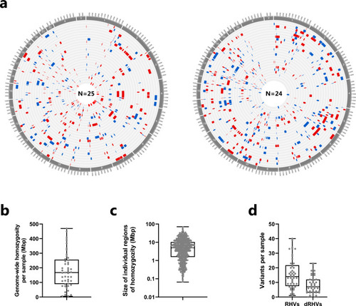
Distribution of rare homozygous variants in CHD patients from consanguineous families. a Genomic regions of homozygosity (ROH) in 49 patients. The genomic localization of ROH, containing at least 50 homozygous variants, is shown with red or blue bars. Each circle represents the genome of one patient. The size of each ROH is shown in (C). a Total size of ROH per patient in mega base-pairs (Mbp). c The size distribution of all 1,167 ROH identified in patients. d The number of rare homozygous variants (RHVs) and damaging RHVs (dRHVs) identified per sample

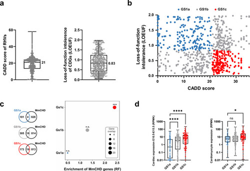
Prioritization of candidate disease genes. a Distribution of CADD scores of RHVs identified in the patients (left) and distribution of loss-of-function intollerence score (LOEUF) in candidate disease genes (CDGs, right). Median values are indicated. b X–Y plot of 678 CDGs containg rare homozygous variants (geneset 1, GS1). The genes are plottet according to CADD score of RHVs identified in each gene (X-axis) and loss-of-function intolerence of the gene (LOEUF, Y-axis). GS1 was divided in three sub-groups. Low-likelihood CDGs (GS1a, blue): CADD < 21, LOEUF > 0.83. Medium likelihood CDGs (GS1b, grey): CADD < 21 and LOEUF ≤ 0.83 or CADD ≥ 21 and LOEUF > 0.83. High-likelihood CDGs (GS1c, red): CADD ≥ 21 and LOEUF ≤ 0.83. c Enrichment of MmCHD genes among three CDG subgroups. Enrichment was calculated by comparing gene-overlap between MmCHD genes and the three sub-groups of GS1. Enrichment is shown as representation factor (RF). A hypergeometric distribution was used to test the significance of the overlaps. d Expression level of the three GS1 subsets in mouse embryonic hearts (left) (Cardoso-Moreira et al. 2019) and in vitro cultures of cardiomyocytes (right) (Wamstad et al. 2012). Difference between medians were determined using ANOVA (Kruskal–Wallis test). Asterisks indicate P values: * P < 0.05, *** P < 0.001, **** P < 0.0001. ns: not significant

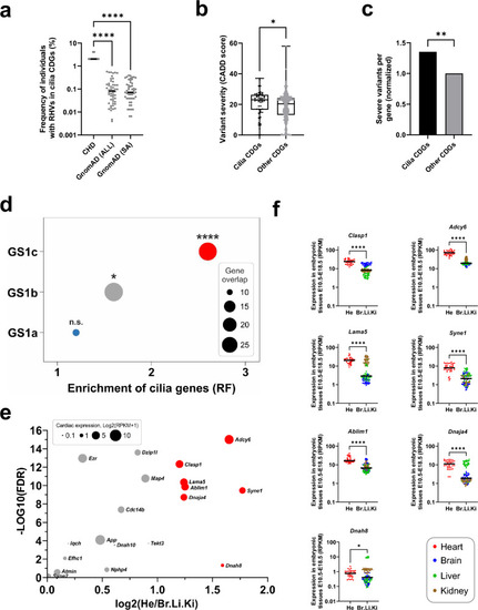
Cilia genes are enriched for rare homozygous variants. a Frequency of individuals with RHVs in 52 cilia CDGs. Frequency per gene in patient cohort (N = 49) was compared with frequency in GnomAD populations. GnomAD (All): Total GnomAD sample (N = 125,748). GnomAD (SA): South Asian GnomAD sub-sample (N = 15,308). Statistic comparison was performed using ANOVA (Kruskal–Wallis test). b Comparison of variant severity of RHVs identified in cilia CDGs (N = 52) and other CDGs (N = 641). Variant severity is indicated as CADD score. Difference between medians were determined using ANOVA (Kruskal–Wallis test). c Normalized comparison of severe RHVs per gene, between cilia CDGs and other CDGs. Severe variants were defined as RHVs with CADD score ≥ 21. Statistical significance of the difference was determined using Fisher’s exact test. d Enrichment of cilia genes among three subsets of GS1. Enrichment was calculated by comparing gene-overlap between CiliaCarta genes (N = 935) and genes within each GS1 subset. Enrichment is shown as representation factor (RF). Hypergeometric statistics was used to test the significance of the overlaps. e Volcano plot of 52 cilia genes showing the differene in gene expression between developing heart (He) and developing brain (Br), Liver (Li) and Kidney (Ki) in mice at E10.5–E18.5 (Cardoso-Moreira et al. 2019). X axis shows the log2 difference between average expression in He and average expression in Br, Li and Ki (only positive values shown). Y axis shows the significance, calculated as –Log10 to the false discovery rate (FDR) (Mann–Whitney U test, adjusted for multiple testing). Significant genes, with log2 difference > 1 is shown with red color. The size of the circle indicate log2 of the average expression of the gene in developing hearts. f Tissue comparative expression of individual genes with fold change > 1. Asterisks indicate P values: *P < 0.05, **P < 0.01, ****P < 0.0001. ns: not significant

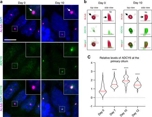
Temporal localization of ADCY6 to primary cilia during cardiomyogenesis. ADCY6 accumulate at primary cilia during differentiation of P19.CL6 cells into cardiomyocytes. a Representative images P19.CL6 cells at day 0 (stem cells) and day 10 (cardiomyocytes). Arrows point to primary cilia. Scale bar, 10 µm. b 3D visualization of representative cilia at day 0 and day 10 of differentiation. Arrow points to the primary cilium. Asterisk marks the ciliary base. c Violin plots of quantification of ADCY6 fluorescence levels at primary cilia during days 0, 7, 10 and 12 of differentiation. Asterisks indicate P values: ****P < 0.0001

Knock-out of ADCY6 cause heart defects in zebrafish. a Bright-field images showing the morphology of 2 dpf uninjected, scramble, adcy6a and adcy6b F0 crispant zebrafish. Scale bars, 0.5 mm. b mRNA expression analysis of myl7 in 2dpf crispant hearts. Upper panels of (i) and (ii) imaged from control larvae. Lower panels imaged from (i) adcy6a and (ii) adcy6b crispants. Scale bars, 50 µm. c, d Quantification of cardiac defects observed in 2 dpf cadcy6a and dadcy6b crispants upon mRNA expression analysis of myl7. ef Proportion of heart phenotypes observed in eadcy6a and fadcy6b crispants. Numbers central within bars indicate number of larvae in each classification. gh Normalized heart rate measurements in beats per minute (bpm) analyzed at 2 dpf in gadcy6a and hadcy6b crispants. Two-way ANOVA (c, d), ordinary one-way ANOVA (g) and Kruskal–Wallis test (h) used for statistical analysis. Asterisk indicate P values: ***p < 0.001. n.s.: not significant

Acknowledgments
This image is the copyrighted work of the attributed author or publisher, and ZFIN has permission only to display this image to its users. Additional permissions should be obtained from the applicable author or publisher of the image. Full text @ Hum. Genet.