
- Title
-
Localization of Piezo 1 and Piezo 2 in Lateral Line System and Inner Ear of Zebrafish (Danio rerio)
- Authors
- Aragona, M., Mhalhel, K., Pansera, L., Montalbano, G., Guerrera, M.C., Levanti, M., Laur�, R., Abbate, F., Vega, J.A., German�, A.
- Source
- Full text @ Int. J. Mol. Sci.

Transmission electron micrograph of a transverse section of an adult zebrafish (D. rerio) canal neuromast. Heterogeneity among sensory hair cells (HCs) can be observed. (a) A HC with a light cytoplasm (green star) and HCs with a more dense, strongly stained cytoplasm (asterisks), both with sparce heterochromatin nucleus (n), vesicle (v), and numerous electron-dense mitochondria (m), are evident. Note an HC showing a group of stereocilia with a typical staircase arrangement (st) in addition to a detached kinocilium (K) and its cross-sections (green arrows). Junction complexes between an HC and a support cell (sc) are indicated by a red inset. At the basal pole of the sensory hair cells, afferent (yellow inset) and efferent (thick arrow) synapses are visible. The occurrence of maturing HCs close to the apical surface is characterized by a peculiar crypt-like rounded space (arrowheads), with stereocilia (s). A maturing volcano-like HC, already reaching the neuromast lumen, with a peculiar depression (broken-arrow) and with the stereocilia surface projecting from the cell surface (s) to the lumen is identified. Elongated scs underneath or close to the HC, sending thin cytoplasmic projections apically, are evident. Note some microvilli (thin arrows) on the top of the sc. (b) Higher magnification of the basal pole of a sensory hair cell. Afferent synapse (aa) characterized by a classical pre-synaptic body (arrowhead) and a post-synaptic side with a clear cytoplasm and mitochondria (m) are visible. (c) Higher magnification of the apical surface of an HC and an sc. Zonula occludens at the apical surface (broken arrow) and more basally desmosomal-like junctions between the HC and sc are clearly visible (gallon arrow). Mitochondria (m). (d) Numerous HCs with dense (asterisk) and lighter cytoplasm (green stars) placed close to the neuromast apical surface. The occurrence of stereocilia (s) in the apical part of the HC. The occurrence of a maturing pear-shaped HC (red star) extending to the apical surface from the basal lamina to the apical surface of the neuromast. Note at the apical pole a peculiar, rounded space with some stereocilia inside (arrowhead). Note the distribution of heterochromatin in the nucleus (n). Mitochondria (m). Afferent synapse (arrow). sc with cytoplasmic projections (in between arrows) separating two adjacent HCs and extending to the apical surface. (a,d) 5000�, (b,c) 10,000�. (e) A graphical representation of the cell subpopulations of the neuromast sensory epithelium: mantle cells (blue), maturing crypt-like HC (red cell), sc (purple cells), hair cells with dense cytoplasm (green cell), hair cells with light cytoplasm (light green cell), maturing volcano-like HC (orange) (Laur� et al. [29] modified). |
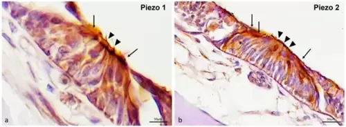
Piezo 1 and Piezo 2 immunoreactivity in the free neuromasts of the zebrafish (D. rerio) lateral line system. (a) Sensory hair cells (arrowheads) and mantle cells (arrows); Piezo 1 immunolabeled. (b) Sensory hair cells (arrowheads) and mantle cells (arrows); Piezo 2 immunopositive. Magnification 40�. EXPRESSION / LABELING:
|

Piezo 1, Piezo 2 immunoreactivity in the canal neuromasts of the zebrafish (D. rerio) lateral line system. (a) Piezo 1 immunoreaction in sensory hair sensory cells (arrows), maturing sensory hair cells (blue arrow heads), nerve (gallon arrow), and bone (asterisk) was observed; (b) hair sensory cells (arrows), maturing sensory hair cells (blue arrow heads), nerve (gallon arrow) s100 immunopositive; (c) Piezo 1/s100p double-stained in sensory hair cells (arrows) and maturing sensory hair cells (blue arrow heads) was observed. Moreover, the nerve (gallon arrow) showed a lower double immunoreaction; (d) Piezo 2 immunoreaction in sensory hair sensory cells (arrows), maturing sensory hair cells (blue arrow heads), and bone (asterisk) was observed; (e) sensory hair sensory cells (arrows) and maturing sensory hair cells (blue arrow heads) s100 immunoreactive; (f) Piezo 2/s100 double-stained in sensory hair sensory cells (arrows) and maturing sensory hair cells (blue arrow heads). Magnification 40�. EXPRESSION / LABELING:
|

Piezo 1 immunoreactivity in the inner ear of zebrafish (D. rerio). (a) The ciliate sensory cells (arrows) in the crista ampullaris and nerve (gallon arrows) that reach the crista ampullaris Piezo 1 immunoreactive; (b) the ciliate sensory cells (arrows) in the macula of the lagena and nerve (gallon arrows) that reach the macula of the lagena Piezo 1 immunopositive; (c) ganglion of the eighth cranial nerve Piezo 1 immunostained; (d) sensory ciliate cells (arrows) of the crista ampullaris and nerve (arrows per gallon) that reach the crista ampullaris s100p immunoreactive; (e) sensory ciliate cells (arrows) of the crista ampullaris and nerve (gallon arrows) reaching the crista ampullaris Piezo 1 immunopositive; (f) ciliate sensory cells (arrows) of the crista ampullaris and nerve reaching the crista ampullaris (gallon arrows) Piezo 1 and s100p double-labeled; (g) neuron (arrows) of ganglion of the eighth cranial nerve s100p immunostained and (h) Piezo 1 immunoreactive; (i) s100p/Piezo 1 double-labeled in some neurons of the eighth cranial nerve ganglion. Magnification 40�. EXPRESSION / LABELING:
|

Piezo 1 immunoreactivity in the inner ear of zebrafish (D. rerio). (a) Sensory hair cells (arrows) and nerve (gallon arrows) of the ampullate crest BDNF immunoreactive; (b) sensory hair cells (arrows) and nerve (gallon arrows) of the crista ampullaris Piezo 1 immunopositive; (c) Piezo 1/BDNF double-staining in some sensory hair cells (arrows) and nerve (gallon arrows) in the crista ampullaris; (d) sensory cells (arrows) in the macula of the utricle BDNF immunoreactive; (e) sensory cells (arrows) in the macula of the utricle Piezo 1 immunopositive; (f) sensory cells (arrows) in the macula of the utricle BDNF/Piezo 1 double-stained; (g) sensory cells (arrows) and nerve (gallon arrows) in the macula of lagena BDNF immunoreactive; (h) sensory cells (arrows) and nerve (gallon arrows) in the macula of the lagena Piezo 1 immunostained; (i) sensory cells (arrows) and nerve (gallon arrows) in the macula of the lagena Piezo 1/BDNF double-labeled; (j) sensory cells (arrows) and nerve (gallon arrows) in the macula sacculus BDNF immunoreactive; (k) sensory cells (arrows) and nerve (gallon arrows) in the macula sacculus Piezo 1 immunopositive; (l) sensory cells (arrows) and nerve (gallon arrows) in the macula of sacculus Piezo 1/BDNF double-stained. Magnification 40�. EXPRESSION / LABELING:
|
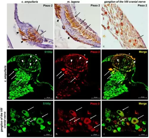
Piezo 2 immunoreactivity in the zebrafish (D. rerio) inner ear and ganglion of the eighth cranial nerve. (a) The ciliate sensory cells (arrows) in the crista ampullaris and nerve (gallon arrows) that reach the crista ampullaris Piezo 2 immunoreactive; (b) the ciliate sensory cells (arrows) in the macula of the lagena and nerve (gallon arrows) that reach this macula Piezo 2 immunopositive; (c) ganglion of the eighth cranial nerve Piezo 2 immunostained; (d) sensory ciliate cells (arrowheads) of the crista ampullaris, cilia (asterisk), and nerve (arrows) reaching the crista ampullaris immunoreactive to s100p; (e) sensory ciliate cells (arrowheads) of the crystal ampullaris, cilia (asterisk), and nerve (arrows) reaching the crista ampullaris Piezo 2 immunopositive; (f) ciliate sensory cells (arrowheads) of crista ampullaris, cilia (asterisk), and nerve reaching the crystal ampullaris (arrows) Piezo 2 and s100p double-labeled; (g) neuron (arrows) of ganglion of the eighth cranial nerve s100p (h) and Piezo 2 immunostained; (i) s100p/Piezo 2 double-labeled in some neurons of the eighth cranial nerve ganglion. Magnification 40�. EXPRESSION / LABELING:
|

Piezo 2 immunoreactivity in the zebrafish (D. rerio) inner ear. (a) Sensory hair cells (arrows), cilia (asterisk), and nerve (gallon arrow) that reach the ampullate crest BDNF immunoreactive; (b) sensory ciliate cells (arrows), cilia (asterisk), and nerve (gallon arrow) reaching the Piezo 2 immunopositive ampullate crest; (c) Piezo 1/BDNF double-staining in some sensory hair cells (arrows), cilia (asterisk), and nerve (gallon arrow) that reaches the ampullate crest; (d) sensory cells (arrows) in the macula of the utricle BDNF immunoreactive; (e) sensory cells (arrows) in the macula of the utricle Piezo 2 immunopositive; (f) sensory cells (arrows) in the macula of the utricle BDNF/Piezo 2 double-stained; (g) sensory cells (arrows) and nerve (gallon arrows) in the macula of the lagena BDNF immunoreactive; (h) sensory cells (arrows) and nerve (gallon arrows) in the macula Piezo 2 immunostained; (i) sensory cells (arrows) and nerve (gallon arrows) in the macula of the lagena Piezo 1/BDNF double-labeled; (j) sensory cells (arrows) and nerve (gallon arrows) in the macula of the sacculus immunoreactive to BDNF; (k) sensory cells (arrows) and nerve (gallon arrows) in the macula of the sacculus immunopositive to Piezo 2; (l) sensory cells (arrows) and nerve (gallon arrows) in the macula of the sacculus Piezo 2/BDNF double-stained. Magnification 40�. EXPRESSION / LABELING:
|

Graphical representation of immunoreactive cell counts: hair sensory cells and maturing hair sensory cells, mantle cells, and nerve in the neuromast epithelium labeled by Piezo 1, Piezo 2, and s100p; sensory hair cells and nerve of inner ear crista ampullaris and maculae epithelium immunolabeled by Piezo 1, Piezo 2, BDNF, and s100p; neurons of VIII cranial nerve immunostained with Piezo 1, Piezo 2, and s100p. The statistical analysis shows a different expression pattern of the investigated protein in different cellular subpopulations. N�: mean of cells immunopositive to Piezo 1, Piezo 2, BDNF, and s100p. The significant difference was assessed for p < 0.05. |