- Title
-
Expression and Roles of Teneurins in Zebrafish
- Authors
- Cheung, A., Trevers, K.E., Reyes-Corral, M., Antinucci, P., Hindges, R.
- Source
- Full text @ Front. Neurosci.
|
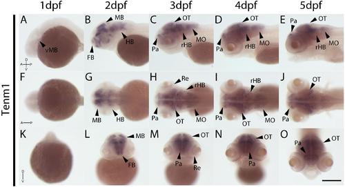
Wholemount expression of tenm1 during zebrafish development. Tenm1 expression during 1–5 dpf shown from lateral (A–E), dorsal (F–J) and frontal (K–O)perspectives. A, Anterior; D, Dorsal; FB, Forebrain; HB, Hindbrain; MB, Midbrain; MO, Medulla Oblongata; OT, Optic Tectum; Pa, Pallium; P, Posterior; Re, Retina; rHB, rostral Hindbrain; V, Ventral; vMB, ventral Midbrain. Scale bar in all panels = 250 μm.
|
|
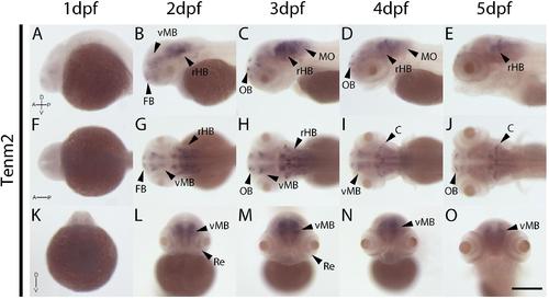
Wholemount expression of tenm2 during zebrafish development. Tenm2 expression during 1–5 dpf shown from lateral (A–E), dorsal (F–J) and frontal (K–O)perspectives. A, Anterior; C, Cerebellum; D, Dorsal; FB, Forebrain; MO, Medulla Oblongata; OB, Olfactory Bulb; P, Posterior; Re, Retina; rHB, rostral Hindbrain; V, Ventral; vMB, ventral Midbrain. Scale bar in all panels = 250 μm.
|
|
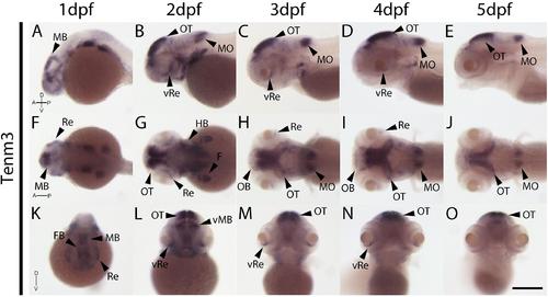
Wholemount expression of tenm3 during zebrafish development. Tenm3 expression during 1–5 dpf from lateral (A–E), dorsal (F–J) and frontal (K–O)perspectives. A, Anterior; D, Dorsal; F, Fin; FB, Forebrain; HB, Hindbrain; MB, Midbrain; MO, Medulla Oblongata; OB, Olfactory Bulb; OT, Optic Tectum; P, Posterior; Re, Retina; V, Ventral; vRe, ventral Retina. Scale bar in all panels = 250 μm.
|
|
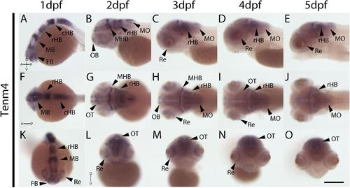
Wholemount expression of tenm4 during zebrafish development. Tenm4 expression during 1–5 dpf from lateral (A–E), dorsal (F–J) and frontal (K–O)perspectives. A, Anterior; cHB, caudal Hindbrain; D, Dorsal; FB, Forebrain; MB, Midbrain; MHB, Mid-Hindbrain Boundary; MO, Medulla Oblongata; OB, Olfactory Bulb; OT, Optic Tectum; P, Posterior; rHB, rostral Hindbrain; Re, Retina; V, Ventral. Scale bar in all panels = 250 μm.
|
|
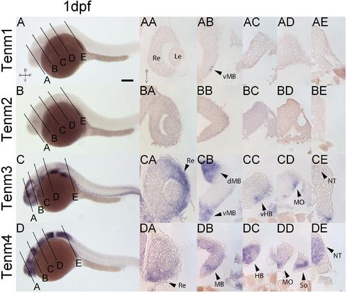
Comparing tenm1–4 expression at 1 dpf. Transverse sections through the retina (A), midbrain (B), rostral hindbrain (C), caudal hindbrain (D) and spinal cord (E) of 1 dpf zebrafish expressing tenm1–4. A, Anterior; D, Dorsal; dMB, dorsal Midbrain; HB, Hindbrain; Le, Lens; MB, Midbrain; MO, Medulla Oblongata; NT, Neural Tube; P, Posterior; Re, Retina; So, Somites; vMB, ventral Midbrain; V, Ventral; vHB, ventral Hindbrain. Scale bar = 150 μm in A–D; 30 μm in all other panels.
EXPRESSION / LABELING:
|
|
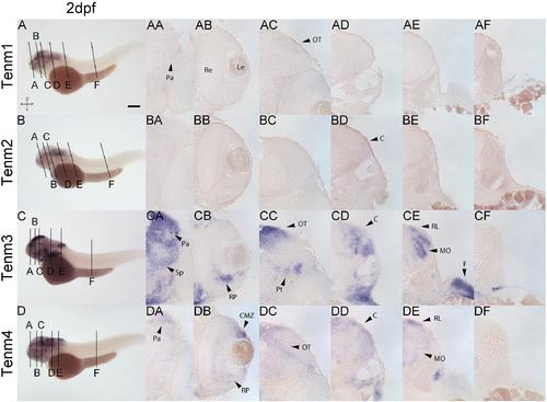
Comparing tenm1–4 expression at 2 dpf. Transverse sections through the forebrain (A), retina (B), midbrain (C), rostral hindbrain (D), caudal hindbrain (E)and spinal cord (F) of 2 dpf zebrafish expressing tenm1–4. A, Anterior; C, Cerebellum; CMZ, Ciliary Marginal Zone; D, Dorsal; F, Fin; Le, lens; MO, Medulla Oblongata; OT, Optic Tectum; Pa, Pallium; P, Posterior; Pt, Pretectum; Re, Retina; RL, Rhombic Lip; Rp, Retinal Progenitors; Sp, Subpallium; V, Ventral. Scale bar = 250 μm in A–D; 30 μm in all other panels.
EXPRESSION / LABELING:
|
|
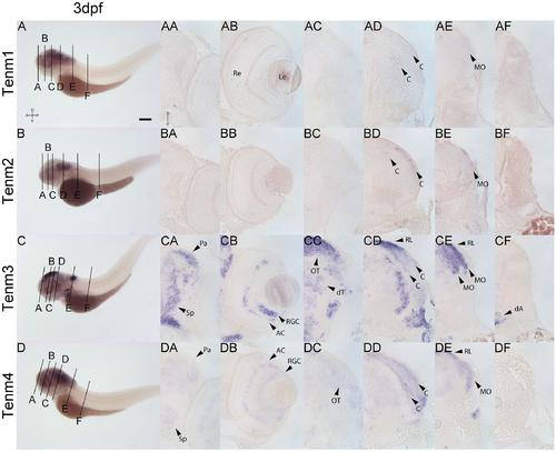
Comparing tenm1–4 expression at 3 dpf. Transverse sections through the forebrain (A), retina (B), midbrain (C), rostral hindbrain (D), caudal hindbrain (E)and spinal cord (F) of 3 dpf zebrafish expressing tenm1–4. A, Anterior; AC, Amacrine Cells; C, Cerebellum; dA, dorsal Aorta; D, Dorsal; dT, dorsal Thalamus; Le, Lens; MO, Medulla Oblongata; OT, Optic Tectum; Pa, Pallium; P, Posterior; Re, Retina; RGC, Retinal Ganglion Cells; RL, Rhombic Lip; Sp, Subpallium; V, Ventral. Scale bar = 250 μm in A–D; 30 μm in all other panels.
EXPRESSION / LABELING:
|
|
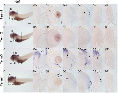
Comparing tenm1–4 expression at 4 dpf. Transverse sections through the forebrain (A), retina (B), midbrain (C), rostral hindbrain (D), caudal hindbrain (E)and spinal cord (F) of 4 dpf zebrafish expressing tenm1–4. A, Anterior; AC, Amacrine Cells; C, Cerebellum; D, Dorsal; Le, Lens; MO, Medulla Oblongata; OT, Optic Tectum; Pa, Pallium; P, Posterior; Re, Retina; RGC, Retinal Ganglion Cells; RL, Rhombic Lip; V, Ventral. Scale bar = 250 μm in A–D; 30 μm in all other panels.
EXPRESSION / LABELING:
|
|
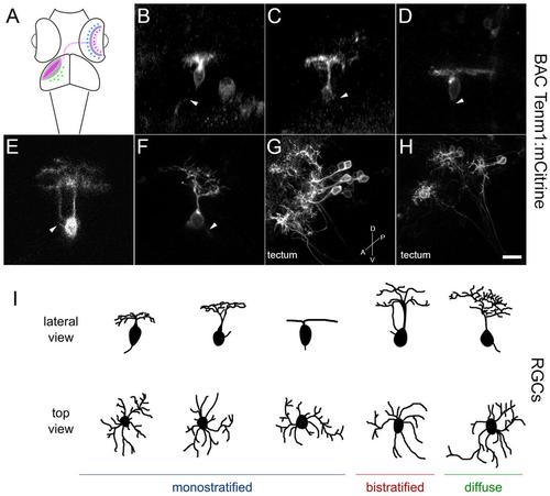
Tenm1-positive cells in the zebrafish visual system in tenm1:mCitrine transient transgenic fish at 3 dpf. Schematic of amacrine cells (blue), RGCs (magenta) and tectal cells (green) in the zebrafish visual system (A). Lateral view of representative RGCs with monostratified (B–D), bistratified (E) and diffuse (F) dendritic arbors. RGC axons are marked by arrowheads. Tenm1-positive tectal cells (G–H). All images represent maximum intensity projections of ∼30 μm confocal z-stacks. Scale bar 10 μm (B–F), 30 μm (G) and 20 μm (H). Schematic representation of tenm1-positive RGC morphology based on representative RGCs imaged in tenm1:mCitrine transient transgenic fish at 3 dpf (I).
|
|
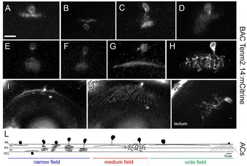
Tenm2.14-positive cells in the zebrafish visual system in tenm2.14:mCitrine transient transgenic fish at 4 and 5 dpf. Lateral view of representative narrow field (A–F), medium field (G–H) and wide field (I) amacrine cells. Top view of a wide field amacrine cell showing ample dendritic coverage of the retina (J). Lateral view of a tectal cell (K). All images represent maximum intensity projections of ∼30 μm confocal z-stacks. Scale bar 10 μm (A–H), 20 μm (I–J) and 15 μm (K). Schematic representation of tenm2.14-positive amacrine cell morphology based on representative cells imaged in tenm2.14:mCitrine transient transgenic fish at 4 and 5 dpf (L).
|

