- Title
-
The Dual Functional Reflecting Iris of the Zebrafish
- Authors
- Gur, D., Nicolas, J.D., Brumfeld, V., Bar-Elli, O., Oron, D., Levkowitz, G.
- Source
- Full text @ Adv Sci (Weinh)
|
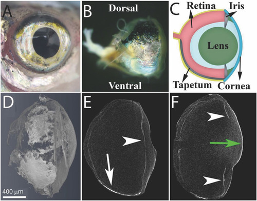
The anatomy of the zebrafish eye. A) A top view of the zebrafish eye showing the silvery iris. B) A side view of the fish eye showing the tapetum lucidum, a silvery layer, located at the ventral side of the eyeball and a pigmented layer visible at the dorsal side of the eye. C) A schematic representation of the eye showing the cornea (blue), lens (green), and retina (light red), as well as the guanine?based reflecting layers, the iris argenta and the tapetum lucidum (silver). D?F) Micro?CT images of the zebrafish eye: a volume rendering of the whole eye, a sagittal view of the lower part of the eye, and a sagittal view of the center of the eye. Arrowheads indicate the iris argentum, white arrow indicates the tapetum lucidum, and green arrow indicates the cornea. Scale bars: 400 Ám.
|
|
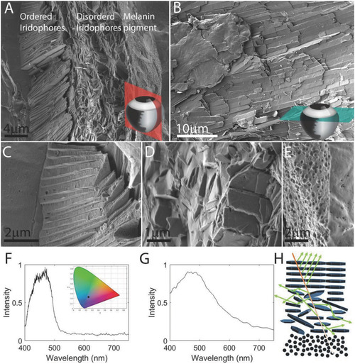
The ultrastructure and optical properties of the iris. A) A coronal section through the iris showing three distinct layers: (i) ordered iridophores, (ii) disordered iridophores, and (iii) a pigmented layer. The section was taken from the periphery of the eye where the iris is curved and, thus, the crystals in the ordered layer are tilted. B) A top view of the iris obtained by a transverse section through the eye shows the elongated guanine crystals packed one next to the other, perfectly tiling the surface of the iris. The anatomical location of sections is illustrated in the bottom?right corners in (A) and (B). C?E) Higher magnifications of the different iris layers: ordered iridophores, disordered iridophores, and pigmented layer. A?E) Cryo?SEM images of high?pressure frozen, freeze?fractured iris. F) Graph showing measured reflectance from the iris, revealing a broad peak centered around 450 nm. Inset shows the color of reflected light (black dot) plotted on a 1931 CIE chromaticity space diagram. G) Simulated reflectance of the ordered layer calculated based on the data in high?resolution cryo?SEM images. H) A sketch illustrating the different paths of light traveling through the multilayered iris. Scale bars: 4 Ám (A), 10 Ám (B), 2 Ám (C,E), 1 Ám (D).
|
|
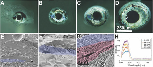
Iris development. Iris morphology, ultrastructural and optical properties were examined during development at 7 (A,E), 14 (B,F), 21 (C,G), and 28 (D) dpf. A?D) Light microscope images of the larva iris at different stages. E?G) Cryo?SEM images of high?pressure frozen, freeze?fractured larva iris show the ultrastructure of the developing iris. Whereas at 7 and 14 dpf only the ordered layer is seen, by 21 dpf a second, disordered layer starts to appear. Ordered iridophores (purple) and disordered iridophores (light red) are shown in pseudo?colors. H) Graph showing measured reflectance of irises from 7, 14, 21, and 28 dpf larvae, revealing a peak reflectance centered around 450 nm. Scale bars: 250 Ám (A?D), 4 Ám (E?G).
|
|
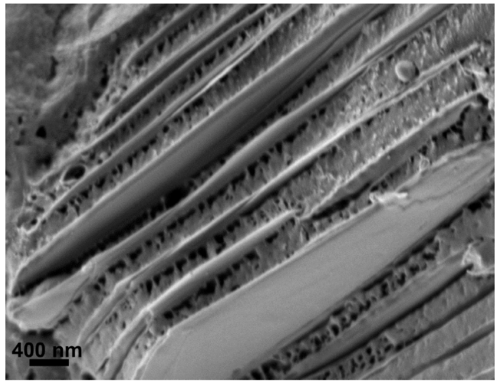
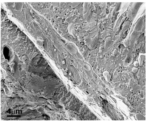
Cryo-SEM image of the ordered iridophore layer in an adult zebrafish iris. Image of a freeze-fracture adult iris shows the parallel stack of thin guanine crystals separated by thicker cytoplasm spacings. |
|
Cryo-SEM image of the tapetum layer in a 21dpf zebrafish eye. Image of a freeze-fracture adult eye shows that the tapetum layer is composed only from one ordered layer of iridophores. |
|
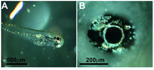
Light microscopy images of 3 dpf zebrafish larvae. (A) A complete larva. (B) Magnification of the eye. Already at this stage, the iridophores in the larval iris are clearly visible. The iridophores surrounding the lens seem to be the first to form. Scale bars: 500 ?m (A), 200 ?m (B). |
|
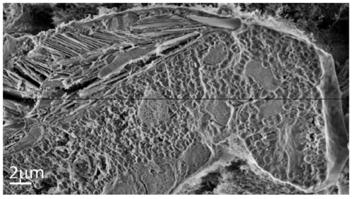
Cryo-SEM image of the zebrafish iris at 21 dpf. Image of a freeze-fractured iris shows the area adjacent to the lens, where two layers of iridophores are visible. Scale bar: 2 ?m. Figure |
|
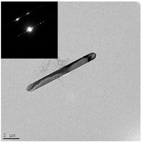
TEM images of a single guanine crystal. A TEM image showing a single crystal extracted from the zebrafish iris and its electron diffraction (inset). The electron diffraction correspond to anhydrous ?-guanine. |
|
Light microscope images of the outer iridophores surrounding the eye. This layer extends to the point where the eye is protruding out from the fish head. A) Ventral view of the eye. B) A side view from posterior to anterior. C) A dorsal view of the eye. |
|
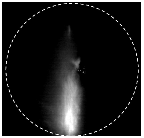
Fourier transform image of an adult zebrafish iris reflectance. A microscopic image showing the highly directional reflection of an adult zebrafish iris. |

