
- Title
-
Testing of therapies in a novel nebulin nemaline myopathy model demonstrate a lack of efficacy
- Authors
- Sztal, T.E., McKaige, E.A., Williams, C., Oorschot, V., Ramm, G., Bryson-Richardson, R.J.
- Source
- Full text @ Acta Neuropathol Commun
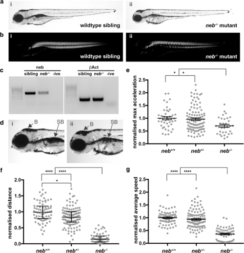
Characterization of the neb (sa906) mutant zebrafish strain. a & b) At 4 dpf neb?/? zebrafish (aii) appear smaller in size and (bii) display a loss of birefringence compared to their wildtype siblings (ai & bi). c) RT-PCR analysis in and neb?/? mutant embryos at 2 dpf shows a reduction in neb mRNA levels compared to wildtype siblings (sibling). ?Act was used as a positive control. d) ii) neb?/? mutants display a smaller eye, brain region (B) and deflated swim bladder (SB) compared to their i) wildtype siblings. e) Quantification of the maximum acceleration recorded from touch-evoked response assays of neb?/? fish compared to wildtype siblings at 2 dpf. Error bars represent mean�SEM for three independent experiments (n =?11,9,12 neb ?/? , 55,25,32 neb +/? , 18,13,12 neb +/+ zebrafish per experiment), **p <?0.01. f & g) Quantification of the normalized (f) distance and (g) speed travelled by neb?/? mutants compared to wildtype siblings at 6 dpf. For f) error bars represent median�interquartile range for three independent experiments (for n =?19,23,19 neb ?/? , 41,42,36 neb +/? , 31,20,21 neb +/+ zebrafish). For g) error bars represent mean�SEM range (for n?=?19,23,14 neb ?/? , 41,42,36 neb +/? , 30,20,21 neb +/+ zebrafish per experiment). *p <?0.5, **** p <?0.001 |

Characterisation of skeletal muscle pathology in neb?/? fish. a Gomori trichome staining of neb?/? skeletal muscle sections reveal the presence of dark regions (arrows) throughout the muscle indicative of nemaline bodies not observed in neb +/+ fish. Nuclei (arrowhead) are evenly organized in neb +/+ , however, appear disorganized in neb?/? fish. b Quantification of normalized fiber area from Gomori trichome stained sections in neb?/? (n?=?23 fibers) compared to neb+/+ fish (n?=?21 fibers). Error bars represent mean�SD, *** p <?0.001. c neb?/? mutants exhibit F-actin (red) and Actinin2 (green) positive aggregates at the myosepta (arrowheads) (and zoomed inset) compared to wildtype siblings at 2 dpf |

Examination of neb?/? skeletal muscle by electron microscopy. a) neb ?/? mutant skeletal muscles display (i, iv) thickened Z-disks (arrows), (ii) fiber breakage (asterisks), (iii) accumulations of nemaline bodies and (iv) disruption of sarcomeric structures that are not observed in b) neb +/+ wildtype siblings PHENOTYPE:
|
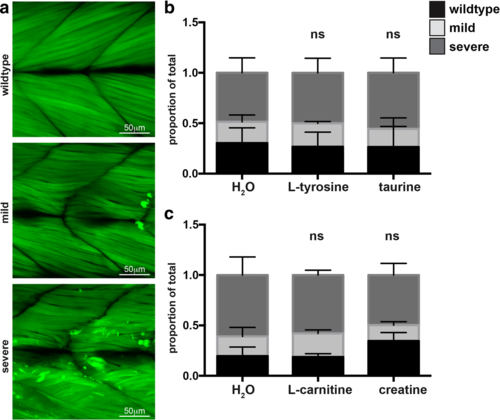
Quantification of the phenotypic severity of neb ?/? mutants at 6 dpf. Quantification of the phenotypic severity of Tg(neb ?/? ; Lifeact-eGFP) fish at 6 dpf supplemented with either L-tyrosine, taurine, L-carnitine, creatine, or water (H2O). a Phenotypes were scored as either wildtype, mild (less than five Lifeact-eGFP positive aggregates at the myosepta or a mild disruption of muscle fibres), or severe (severely disorganised fibres or an accumulation of five or more Lifeact-eGFP positive aggregates within the muscle cell). b Quantification of the phenotypic severity of Tg(neb ?/? ; Lifeact-eGFP) fish supplemented with either L-tyrosine, taurine, or water. c Quantification of the phenotypic severity of Tg(neb ?/? ; Lifeact-eGFP) fish supplemented with either L-carnitine, creatine, or water (H2O). b & c Error bars represent mean�SEM for three independent experiments. For b) n =?6,8,7 Tg(neb ?/? ; Lifeact-eGFP) for L-tyrosine, n?=?11,5,11 Tg(neb ?/? ; Lifeact-eGFP) for taurine and n?=?9,8,10 Tg(neb ?/? ; Lifeact-eGFP) for water. For c) n?=?8,10,4 Tg(neb ?/? ; Lifeact-eGFP) for L-carnitine, n?=?6,8,3 Tg(neb ?/? ; Lifeact-eGFP) for creatine, and n =?10,9,5 Tg(neb ?/? ; Lifeact-eGFP) for water per experiment). ns?=?not significant |
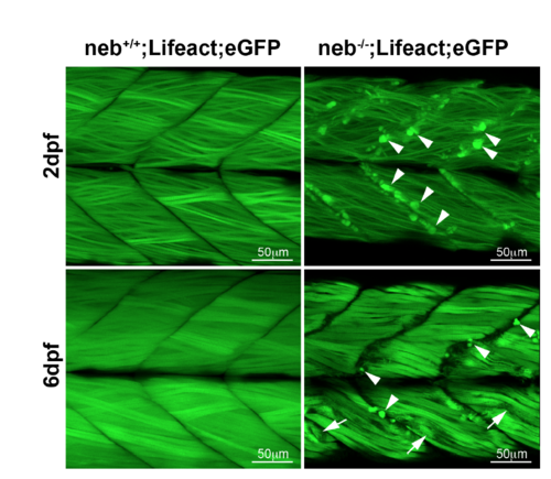
Characterisation of Tg(neb-/-; Lifeact-eGFP) fish. Tg(neb-/-; Lifeact-eGFP) fish show an accumulation of Lifeact-eGFP at the myosepta (arrowheads) at both 2 dpf and 6 dpf as well as regions of disorxganized muscle fibers (arrows) at 6 dpf that are not observed in Tg(neb+/+; Lifeact-eGFP) siblings. |
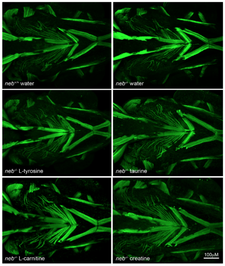
Characterisation of facial muscles in Tg(neb-/-; Lifeact-eGFP) fish and wild type siblings at 6 dpf. Maximum projection images of Tg(neb-/-; Lifeact-eGFP) fish supplemented with water, tyrosine, taurine, L-carnitine of creatine show no difference in the appearance of facial muscles to Tg(neb+/+; Lifeact-eGFP) siblings supplemented with water. |