- Title
-
Conversion of the thymus into a bipotent lymphoid organ by replacement of FOXN1 with its paralog, FOXN4
- Authors
- Swann, J.B., Weyn, A., Nagakubo, D., Bleul, C.C., Toyoda, A., Happe, C., Netuschil, N., Hess, I., Haas-Assenbaum, A., Taniguchi, Y., Schorpp, M., Boehm, T.
- Source
- Full text @ Cell Rep.
|
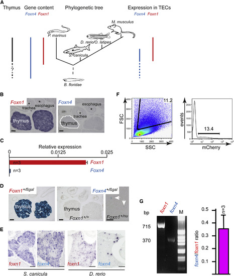
Evolutionary Features of Foxn4 and Foxn1 Genes in Vertebrates (A) Summary of relevant features aligned with a schematic of the chordate phylogenetic tree. The thymus is present in all vertebrates but absent in nonvertebrate chordates. The Foxn1 gene first appears in vertebrates as a paralog of the ancient Foxn4 gene. The expression of Foxn4 in pharyngeal endoderm precedes that of Foxn1. (B) Expression of Foxn1 and Foxn4 in E15.5 mouse thymi as determined by RNA in situ hybridization; scale bar, 100 ?m. (C) Quantitative RT-PCR for Foxn1 and Foxn4 expression in purified adult mouse TECs relative to ?-actin. (D) X-gal staining of E15.5 mouse thymi from Foxn1+/lacZ and Foxn4+/lacZ knockin embryos; note that Foxn4 is expressed at low levels in a small proportion of TECs throughout the thymus, whereas the corresponding analysis of Foxn1 demonstrated the anticipated ubiquitous expression. Note that Foxn1nu represents the original nude allele (Nehls et al., 1994), whereas Foxn1lacZ represents a deleterious knockin allele (Nehls et al., 1996). The nu allele of Foxn1 was used here to avoid interference with the detection of ?-galactosidase activity derived from the Foxn4 knockin allele. Scale bar, 100 ?m. (E) foxn1 and foxn4 RNA expression in the thymus of S. canicula and of D. rerio as determined by RNA in situ hybridization. Scale bars, 100 ?m. (F) Light-scatter profile of dissociated thymic cell populations from adult foxn1:mCherry transgenic zebrafish. Number indicates percentage of live cells in gate (left). Red fluorescence of gated cells (right); the percentage of positive cells is indicated. (G) Presence of foxn1 and foxn4 transcripts (size of cDNA fragments given) in red fluorescent cells from (F) (left). Ratio of foxn4 and foxn1 expression in zebrafish TECs as determined by quantitative RT-PCR (right). In this and all subsequent figures, graphs indicate mean ± SEM, with statistical significance shown (t test) were applicable. See also Figure S1 and Tables S1?S3. EXPRESSION / LABELING:
|

