- Title
-
A Novel Tool to Measure Extracellular Glutamate in the Zebrafish Nervous System In Vivo
- Authors
- MacDonald, R.B., Kashikar, N.D., Lagnado, L., Harris, W.A.
- Source
- Full text @ Zebrafish
|
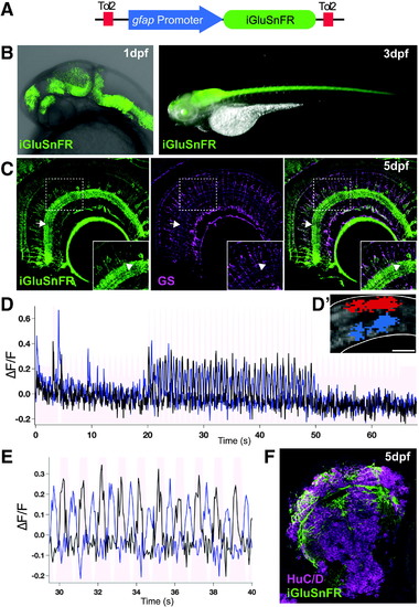
(A) To express iGluSnFR specifically in glial cells, we used the gfap promoter region to generate the Tg(gfap:iGluSnFR) transgenic line. (B) Whole mount fluorescent images of the Tg(gfap:iGluSnFR) embryos showing expression begins at 1 day postfertilization (dpf) in the embryonic and in the maturing nervous system at 3?dpf. (C) Sections showing iGluSnFR expression in the retinal Müller glia cells, labeled with the glutamine synthetase (GS) antibody, up to at least 5?dpf. Inset shows that all GS-positive cells are also iGluSnFR positive in the retina. The iGluSnFR signal is enriched in the inner plexiform layer (arrow) presumably because of the presence of low-level extracellular glutamate in the synaptic layer of the retina. There are occasional GS-negative cells, presumably neurons, expressing iGluSnFR in the inner nuclear layer of the retina (arrowhead). These cells may be labeled because of early activity of the gfap promoter in progenitors during the genesis of neurons, which has been noted previously.5 (D) iGluSnFR in the retina responds to light stimulation as characterized by activity-dependent changes in fluorescence over time in the ON (black plot) and OFF (blue plot) regions of the inner plexiform layer (arrow). (D?) An example of the region of interest used to measure ON (blue) and OFF (red) responses in the inner plexiform layer of the retina. Scale bar denotes 10??m. (E) Zoom of D between 30 and 40?s clearly shows the light-dependent responses in the retina. (F) iGluSnFR is also expressed in the glial cells and neuropil of the brain, but not the neurons (HuC/D positive) in sections at 5?dpf. |

