- Title
-
A Zebrafish Genetic Screen Identifies Neuromedin U as a Regulator of Sleep/Wake States
- Authors
- Chiu, C.N., Rihel, J., Lee, D.A., Singh, C., Mosser, E.A., Chen, S., Sapin, V., Pham, U., Engle, J., Niles, B.J., Montz, C.J., Chakravarthy, S., Zimmerman, S., Salehi-Ashtiani, K., Vidal, M., Schier, A.F., Prober, D.A.
- Source
- Full text @ Neuron
|
A Behavioral Genetic Overexpression Screen (A) The Heat Shock Secretome (hs:Sec) overexpression transgenesis cassette. (B) In the primary screen, single cell zebrafish embryos were co-injected with the hs:Sec plasmid and tol2 transposase mRNA. Embryos were raised to 5 days post-fertilization (dpf) at 28.5°C, a non-permissive temperature for the heat shock promoter. (C) A 1 hr, 37°C heat shock (but not the 28.5°C control) treatment induces expression of a Secretome gene in hs:Sec/+ larvae, as visualized by in situ hybridization (purple stain) in dissected brains. (D) Average activity traces for hs:Hcrt_Dr-injected (n = 32) and control injected (n = 16) larvae. (E) Histogram of all activity indices from the screen, normalized as standard deviations from the mean (Z score). (F) Average activity traces of controls and hs:Pnoc_Hs, a previously identified regulator of sleep/wake behavior in zebrafish. (G) Average activity traces of controls and hs:Nmu_Hs, a newly identified regulator of sleep/wake behavior in zebrafish. (H) Candidate genes were retested as stable transgenic lines. hs:Nmu_Hs/+ larvae generated from a stable transgenic line exhibited increased activity compared to their WT siblings. Line plot values are represented as mean ± SEM. White and black boxes on the x axis of the line plots denote daytime light periods and nighttime dark periods, respectively. Arrows indicate time of heat shock (F-H). *p < 0.05, Mann-Whitney U test. |
|
The Sequence of Nmu and Expression of nmu and nmu receptors Are Conserved in Zebrafish (A) The zebrafish predicted Nmu peptide sequence is well conserved with the human Nmu mature peptide. The C-terminal heptapeptide and critical residues are indicated by the shaded box and asterisks. Amino acids are color coded by type (after Malendowicz et al., 2012): blue, basic; red, acidic; green, non-polar; light green, polar. (B-F) Endogenous expression of nmu in the hypothalamus (single arrow), brainstem (double arrow), and spinal cord (arrowhead) of 24-120 hpf zebrafish. (G and H) Lateral views of nmur1a and nmur2 expression at 48 hpf. (I) nmur1a expression in 5 dpf larvae is restricted to the rostro-ventral hypothalamus. (J-L) Widespread but discrete expression of nmur2 in 5 dpf zebrafish brain, starting from a ventral focal plane (J) and ending in a more dorsal focal plane (L). Note that a prominent population of cells in the hypothalamus (left/rostral arrow in J) does not localize to the zebrafish preoptic nucleus, which is not visible in this focal plane. Also note several discrete clusters of cells in the hindbrain (arrows in L), which are located in the vicinity of brainstem arousal systems such as the locus coeruleus. In (G)-(L), arrows distinguish cellular labeling from diffuse background staining. Scale bars = 100 µm. |
|
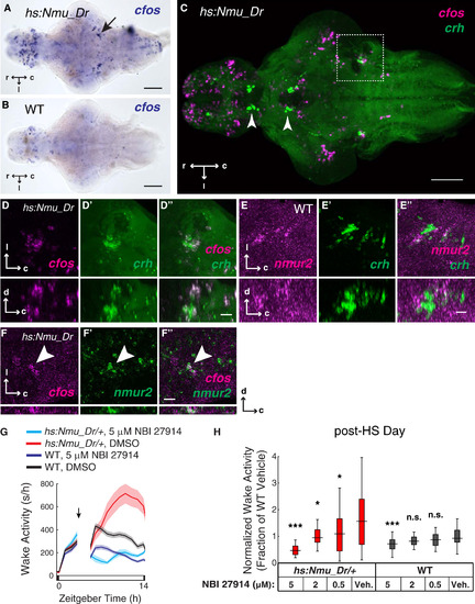
Nmu Overexpression Activates Brainstem crh Neurons and the Nmu Overexpression Locomotor Phenotype Requires Crh Receptor 1 Signaling (A and B) ISH analysis of cfos expression 1 hr after heat-shock induction of Nmu overexpression in a 5 dpf hs:Nmu_Dr/+ transgenic zebrafish brain (A) and in an identically treated non-transgenic sibling (B). Arrow indicates cfos-expressing brainstem populations. (C-D′′) Double fluorescent ISH reveals brainstem colocalization of cfos and crh expression following Nmu overexpression (dotted box is magnified in D), whereas diencephalic crh expression does not colocalize with cfos (arrowheads). The brainstem populations in (C) are presented in separate and overlaid confocal channels at higher magnification dorsal (D-D′′, top) and orthogonal (D-D′′, bottom) views to demonstrate colocalization. (E-E′′) Double fluorescent ISH reveals sparse colocalization of nmur2 and crh expression in the same brainstem populations and in the same views as shown in (D)-(D′′). (F) Following Nmu overexpression, some nmur2-expressing cells colocalize with cfos in the same brainstem populations and in the same views as shown in (E-E′′). Scale bars = 100 µm in (A)-(C) and 20 µm in (D)-(F). Images are maximum intensity projections of optical stack thickness = 300 µm (C), 82 µm (D), 85 µm (E), and 16 µm (F). (G) 5 µM NBI 27914, a Crhr1 antagonist, or vehicle (0.05% DMSO) was applied to hs:Nmu_Dr/+ larvae and their WT siblings immediately after a 1 hr heat shock at 12 p.m. Line plot indicates mean ± SEM. Number of subjects: hs:Nmu_Dr/+, 5 µM NBI 27914 (n = 40), WT, 5 µM NBI 27914 (n = 55), hs:Nmu_Dr/+, DMSO (n = 47), WT, DMSO (n = 49). (H) Additional experiments performed at 2 µM and 0.5 µM NBI 27914 reveal dose-dependent inhibition of the Nmu overexpression phenotype. Data were normalized by dividing the average post-HS day waking activity of each fish by the mean value of within-experiment WT vehicle samples. Normalized values from individual samples across separate experiments were then pooled for analysis and box plots. Number of subjects: hs:Nmu_Dr/+, NBI 27914: 5 µM (n = 40), 2 µM (n = 25), 0.5 µM (n = 35), DMSO (n = 101); WT, NBI 27914: 5 µM (n = 55), 2 µM (n = 23), 0.5 µM (n = 32), DMSO (n = 103). *p < 0.05, **p < 0.01, n.s. p > 0.05, Kruskal-Wallis test followed by the Steel-Dwass test for pairwise multiple comparisons of each drug condition against its same-genotype vehicle control. |
|
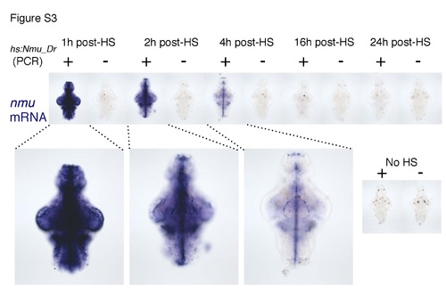
Time course of Nmu overexpression (Related to Figure 3). Larvae from a hs:Nmu_Dr /+ to WT mating were heat shocked for 1 hour at 37oC at 5 dpf and fixed at the indicated times after heat shock. ISH was then performed using an nmu-specific riboprobe on dissected larval brains. Transgenic and WT larvae were subsequently identified by PCR. Ubiquitous nmu overexpression was observed at 1 hour post-heat shock, with reduced levels at 2 and 4 hours post-heat shock. No ectopic transcript was observed at 16 and 24 hours post-heat shock. No ectopic nmu expression was observed in non-heat shocked transgenic brains (No HS, insert). Chromogenic development was stopped at the same time for all samples prior to the visualization of endogenous nmu transcript. While this data shows that ectopic nmu transcript levels are ubiquitously elevated for a few hours after heat shock, the perdurance of Nmu peptide is not known. |
|
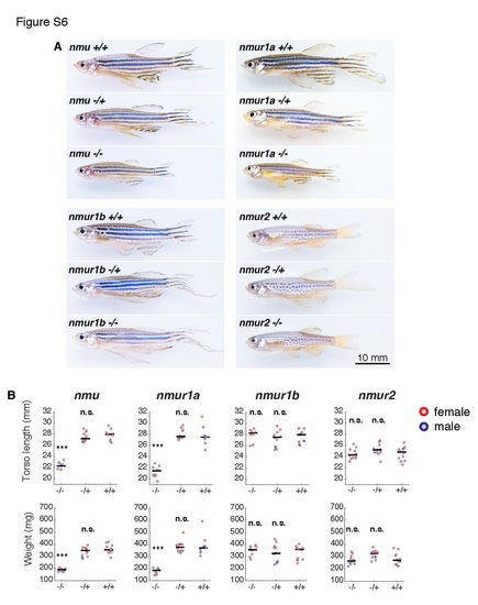
nmu-/- and nmur1a-/- but not nmur1b-/- or nmur2-/- adult zebrafish exhibit defects in size and weight (Related to Figure 5). (A) Representative images of male 20-22 week old adults from each genotype tested. For each mutation, fish from heterozygous mutant matings were raised to adulthood, measured and then genotyped to identify homozygous mutant, heterozygous mutant and WT individuals. All samples are displayed at the same scale (scale bar = 10 mm). (B) Size, as measured from rostral tip to end of torso (fin length not included), and weight of individual fish. Red=female, blue=male. Black horizontal bars indicate median value for each genotype. ***p<0.001, n.s. p>0.05, Kruskal-Wallis test followed by the Steel-Dwass test for pairwise multiple comparisons. PHENOTYPE:
|

Unillustrated author statements |


