- Title
-
The Extreme Anterior Domain Is an Essential Craniofacial Organizer Acting through Kinin-Kallikrein Signaling
- Authors
- Jacox, L., Sindelka, R., Chen, J., Rothman, A., Dickinson, A., Sive, H.
- Source
- Full text @ Cell Rep.
|
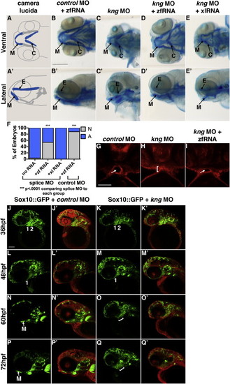
Function of kng in Craniofacial Development Is Conserved in Zebrafish (A and A′) Camera lucida of facial cartilages. E, ethmoid plate; C, ceratohyal cartilage; M, Meckel?s cartilage. (B-E′) kng loss of function using splice morpholinos and rescue with zebrafish (zf) kng mRNA. Embryonic cartilage scored at 5 dpf after Alcian blue staining in three independent experiments. Scale bar, 250 Ám. (B and B′) Control morphants coinjected with mRNA were normal (88% normal, n = 50). (C, C′, E, and E′) kng morphants and kng morphants coinjected with 200 ng Xenopus kng mRNA showed abnormal facial cartilage. Meckel?s cartilage was truncated, boxy, and pointed at an abnormal angle. The ceratohyal cartilage was positioned at an abnormal angle, perpendicular to the midline. (kng [C and C′] 3% normal, n = 61; kng mo plus frog mRNA [E and E′] 0% normal, n = 65). (D and D′) kng morphants coinjected with 200 ng zebrafish (zf) mRNA showed partial rescue. Embryos scored as partially rescued if Meckel?s cartilage was longer, more rounded, and pointed dorsally and if ceratohyal cartilage pointed more anteriorly, compared to kng morphants (54% partial rescue, n = 89). (F) Quantification of phenotypes. p values: one-tailed Fisher?s exact test. N, normal or partially rescued phenotype. A, abnormal phenotype. (G-I) Ventral views of mApple-injected embryos at 48 hpf. White arrow: open mouth. White bracket: closed mouth. Scale bar, 100 Ám. (G) Control morphants (100% normal, n = 5). (H) kng splice morphants failed to form open mouths (0% normal, n = 6). (I) kng splice morphants coinjected with 200 ng zf mRNA had open mouths (67% normal, n = 6). (J-Q′) Confocal images of Sox10::GFP zebrafish coninjected with 75 pg mApple and 4 ng control morpholino (100% normal, n = 5) or 4 ng kng splice morpholino (0% normal, n = 5). Paired images of the same embryo show GFP signal alone and GFP with mApple. Numbers indicate pharyngeal arches (PA). Bracket: uncondensed/disorganized cartilage. Lateral view. M, Meckel?s cartilage. Scale bar, 100 Ám. (J-K′) At 36 hpf, NC has migrated into the face of both morphant and control embryos to form first and second PA. (L-M′) At 48 hpf, the first PA has begun to extend under eye to form the lower jaw in both morphant and control embryos. (N and N′) At 60 hpf, first PA has condensed into Meckel?s cartilage in control embryos. (O and O′) At 60 hpf, first PA remains disorganized in morphants and does not condense. (P and P′) At 72 hpf, Meckel?s cartilage is prominent in control embryos. (Q and Q′) At 72 hpf, cartilage of the lower jaw remains disorganized and uncondensed in morphants. EXPRESSION / LABELING:
|
|
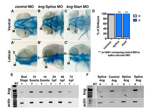
kininogen (kng) is expressed throughout zebrafish craniofacial development and loss of function results in craniofacial cartilage abnormalities, related to Figure 7. (A-C, A′-C′) kng loss of function using splice site and start site morpholinos. Embryonic cartilage was observed at 5dpf using alcian blue staining in three independent experiments. E, Ethmoid plate. C, Ceratohyal cartilage. M, Meckel′s cartilage. Scale bar: 250Ám. (A,A′) Control morpholino injected embryos appeared normal (95% normal, n=43). (B-B′, C-C′) Splice and start morphants showed abnormal facial cartilage. Meckel′s cartilage is truncated and lies at an abnormal angle. The ceratohyal cartilage points at an abnormal angle, perpendicular to the midline. (kng splice morpholino (B,B′) 7% normal, n=95; kng start morpholino (C,C′) 6% normal, n=47). (D) Quantification of morphant phenotypes. P-values: one-tailed Fisher Exact test. (E) Non-quantitative RT-PCR. kng expression is present at bud stage and extends until 72hpf. Expression spans mouth opening at 48hpf and formation of facial cartilage at 72hpf. (F) Non-quantitative RT-PCR. RNA was extracted at 24hpf. Controls yield normal length kng transcripts, while splice morphants yield fewer normal length transcripts and a truncated transcript. Arrow: truncated transcript band. PHENOTYPE:
|


