- Title
-
Identification of a Dopaminergic Enhancer Indicates Complexity in Vertebrate Dopamine Neuron Phenotype Specification
- Authors
- Fujimoto, E., Stevenson, T.J., Chien, C.B., and Bonkowsky, J.L.
- Source
- Full text @ Dev. Biol.
|
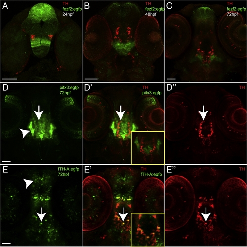
Examples of stable transgenic enhancer lines generated; ventral views, anterior to the top, double-immunohistochemistry for GFP and TH (green and red), confocal z-stacks. Scale bars are 50 μm. (A?C) Time-series of Tg(fezf2:egfp)zc55 expression at 24 hpf, 48 hpf, and 72 hpf, shows non-overlap of TH and GFP expression. (D?D3) Tg(pitx3:egfp)zc50 and (E?E3) Tg(f.TH.A:egfp)zc56 at 72 hpf, shows partial overlap of enhancer expression and TH (arrows), but also wide spread expression of the enhancer in non-TH neurons (arrowheads). Insets in D2 and E2 show single confocal plane of double-labeling, emphasizing the minimal overlap of TH and GFP labeling. EXPRESSION / LABELING:
|
|
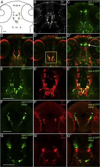
Characterization of Tg(otpb.A:egfp)zc48; confocal images of whole mount embryos double-labeled for GFP and for TH immunohistochemistry, ventral views, anterior to the top. Scale bar is 50 μm. (A) Schematic diagram of TH-positive cell groups in the zebrafish brain at 72 hpf, based on Rink and Wullimann (2002). (B) Confocal z-stack projection of TH immunohistochemistry at 72 hpf in the zebrafish brain. (C) Confocal z-stack projection of GFP immunohistochemistry in Tg(otpb.A:egfp)zc48. (D?D3) Confocal z-stack projections at different dorsal?ventral levels in the brain of Tg(otpb.A:egfp)zc48 at 72 hpf, showing co-expression of TH and GFP in diDA neuron groups 4 and 6, but not groups 1 and 2 (arrow); arrowhead points to the NPO neurons. (E?E3) Magnified views of the region boxed in B3, showing extensive overlap of GFP-positive neurons in the diencephalon with TH expression. (F?F3) Expression at 24 hpf. (G?G3) Expression at 48 hpf. EXPRESSION / LABELING:
|
|
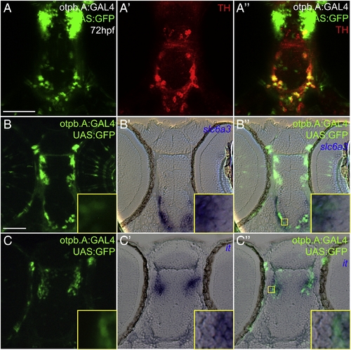
Characterization of otpb.A expression in dopaminergic and neuroendocrine cells. Embryos at 72 hpf, Tg(otpb.A:GAL4)zc57, Tg(UAS:GFP). Scale bar is 50 μm. (A?A3) Confocal z-stack projection of whole-mount embryos double-labeled for GFP and for TH immunohistochemistry, ventral views, anterior to the top. (B) and (C) Plastic horizontal sections of double-labeled Tg(otpb.A:GAL4)zc57; Tg(UAS:GFP) embryos at 72 hpf, anterior to the top. Higher magnification inset showing co-localization is indicated by yellow boxed area and is shown at bottom right of each panel. (B?B3) Labeled for GFP antibody (B) and slc6a3 mRNA (B2), GFP-positive neurons in the diencephalon co-express slc6a3, confirming their identity as dopaminergic. (C?C3) Labeled for GFP antibody (C) and isotocin mRNA (C2), rostral diencephalon GFP-positive NPO neurons co-express GFP and isotocin. EXPRESSION / LABELING:
|
|
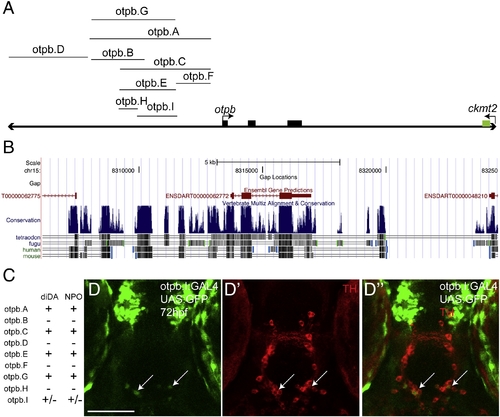
Genomic structure and characterization of otpb genomic locus enhancers. (A) otpb genomic region (not to scale). Coding exons are shown as solid black boxes. DNA fragments tested for enhancer activity are shown. Region pictured is approximately 20 kb; 32-most exon of ckmt2 is shown. (B) Screenshot from the UCSC genome browser (http://genome.ucsc.edu) showing the location of the exons of genes, relative to regions of cross-species conservation (shown as vertical blue lines). Increasing conservation is indicated by increasing height and/or density of the blue lines. Species used to determine the conservation are shown below. (C) Summary table of expression patterns of the different enhancers at 72 hpf in the CNS with respect to expression in the diencephalic dopaminergic neurons (diDA) and neurosecretory preoptic nucleus (NPO). (D?D3) Confocal z-stack projection of whole-mount Tg(otpb.I:GAL4)zc57 embryos at 72 hpf, shown crossed to Tg(UAS:GFP) and double-labeled for GFP (D) and for TH (D2) immunohistochemistry, ventral views, anterior to the top. Arrows point to the sparse diDA neuron expression. EXPRESSION / LABELING:
|
Reprinted from Developmental Biology, 352(2), Fujimoto, E., Stevenson, T.J., Chien, C.B., and Bonkowsky, J.L., Identification of a Dopaminergic Enhancer Indicates Complexity in Vertebrate Dopamine Neuron Phenotype Specification, 393-404, Copyright (2011) with permission from Elsevier. Full text @ Dev. Biol.


