- Title
-
Tbx2 and Tbx3 Regulate the Dynamics of Cell Proliferation during Heart Remodeling
- Authors
- Ribeiro, I., Kawakami, Y., Buscher, D., Raya, A., Rodriguez-Leon, J., Morita, M., Rodriguez Esteban, C., and Izpisua Belmonte, J.C.
- Source
- Full text @ PLoS One
|
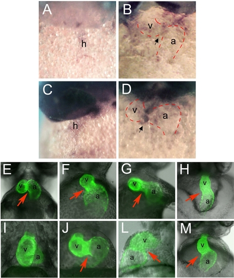
(A?D) Whole mount RNA in situ hybridization of zebrafish tbx3b and tbx2a expression; ventral views show the heart in its maximum extension from the anterior to the posterior pole. Zebrafish tbx3b is expressed throughout the extent of the heart tube at 31 hpf (A) and becomes restricted to the AVC at 42 hpf (B). Zebrafish tbx2a is expressed al low levels throughout the extent of the linear heart tube at 31 hpf (C). At 42 hpf tbx2a transcripts are present in the AVC at high levels (D). Black arrow points to the AVC. (E?M) Ventral views of the heart of a mlc2a::GFP transgenic line that expresses GFP in the myocardium, at 48 hpf (E?H) and 72 hpf (I?M). (E) In wild type 48 hpf embryos, the atrium has moved upward and is positioned at the same anterior-posterior level as the ventricle; (I) later the atrium becomes localized dorsal to the ventricle at 72 hpf. (F, J) Injection of 2.5 ng of tbx3b morpholino into one-cell stage embryos results in delayed heart looping and abnormal AVC. (G, L) Injection of 10 ng of tbx2a MO results in a similar delay in heart looping and an enlargement of the AVC. (H, M) Injection of both tbx3b and tbx2a MOs, at 1.75 and 5 ng respectively, results in the failure to form the AVC constriction and absence of looping. Red arrow indicates the AVC. a, atrium; h, heart; v, ventricle. EXPRESSION / LABELING:
PHENOTYPE:
|
|
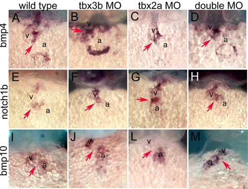
Ventral views of the heart in its maximum extension from the anterior to the posterior pole. (A) bmp4 is expressed in the non-chamber myocardium of the heart, the OFT, the AVC and the IFT. (B, C, D) The expression pattern of bmp4 is normal in 42 hpf tbx3b, tbx2a and double morphants. (E) notch1b is expressed in the whole endocardium of the heart tube, but becomes restricted to the endocardium at the level of the OFT and AVC upon looping. (F, G, H) Expression of notch1b is maintained in the endocardium at the level of the chamber myocardium in 48 hpf tbx3b, tbx2a and double morphants, in contrast to wild type. (I) bmp10 is expressed exclusively in chamber myocardium at 42 hpf (J, L, M) In 42 hpf tbx3b and tbx2a morphants bmp10 is expressed in the chamber myocardium and is downregulated in the AVC, as is the case in wild type, but it is not downregulated from the AVC in double morphants. Red arrow indicates the AVC. a, atrium; h, heart; v, ventricle. EXPRESSION / LABELING:
|
|
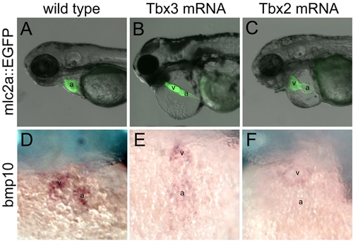
Side views (A?C) and ventral views (D?F) of wild type embryos (A, D) and embryos injected with 100 pg capped mRNA for mouse Tbx3 (B, E) or human TBX2 (C, F). (A, B, C) mlc2a::GFP embryos at 60 hpf; (D, E, F) 48 hpf. (B, C) 44.5% of Tbx3-injected embryos and 60% of Tbx2-injected embryos present a pipe-like heart that lacks chamber growth and looping. (D, E, F) Zebrafish bmp10 is strongly expressed in the chamber myocardium in wild type embryos, but is significantly downregulated from the heart in Tbx3- or Tbx2-injected embryos. a, atrium; v, ventricle. |
|
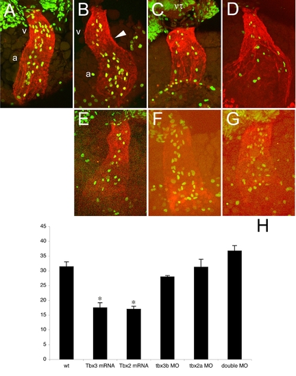
All images represent reconstructions of confocal Z-stack sections imaged on whole embryos at 31hpf (A) and 33 hpf (B?G). (A, B) During the first steps of looping, the pattern of proliferation shifts from homogenous throughout the heart tube (31 hpf, A) to a heterogenous one in which dividing cells are more concentrated in the future chambers (33 hpf, B). (C, D) In Tbx3- (C) and Tbx2- (D) injected embryos at 33 hpf this shift has not occurred and the number of dividing cells was significantly decreased and dividing cells were homogenously distributed (H). (E?G) MO-injected embryos against tbx3b (E), tbx2a (F) or both (G) display the same (E, F) or higher (G) number of proliferating cells than wild type at 33 hpf. However, proliferating cells remain homogenously distributed throughout the heart tube. (H) Histogram showing the average of the total number of BrdU positive cells in the heart of 33 hpf embryos: wt, 31.4±1.661 (n = 5); Tbx3 mRNA, 17.5±1.708 (p<0.001; n = 6); Tbx2 mRNA, 17.0±1.000 (p<0.001; n = 7); tbx3b MO, 28.0±0.408 (n = 4); tbx2a MO, 31.3±2.658 (n = 4); double MO, 36.8±1.797 (n = 4). a, atrium; h, heart; nt, neural tube; v, ventricle. |
|
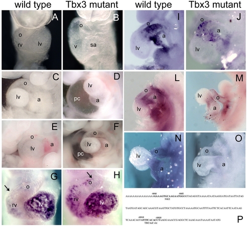
(A, B) Severely affected Tbx3 mutant embryos (B) present a delay in heart looping at E9.5, in which the ventricle is still at the same dorsoventral position as the atrium. (C, D) At E10.5, the most affected Tbx3 mutant embryos display obvious heart defects, including lack of the constriction in the AVC and absence of looping in a swollen pericardiac cavity (D), compared to wild type (C). (E, F) E11.5 Tbx3 null homozygous embryos present a significant delay in heart looping and pericardiac swelling compared to wild type. (G?O) Whole mount in situ hybridization analysis at E9.5; ventral views (G, H) and lateral views (I?O). (G, H) Upon looping initiation, Bmp10 starts to be expressed in the chamber myocardium. However, Bmp10 was ectopically expressed in the non-chamber myocardium of the heart of Tbx3 mutant embryos (arrow in G, H). (I, J) Bmp2 is expressed in the AVC myocardium at E9.5 and was not altered in Tbx3 mutant embryos. (L, M) TGFβ2 is normally expressed in the non-chamber myocardium of the looping heart. In Tbx3 mutant embryos, expression of TGFβ2 is downregulated in the heart. (N, O) Smad6 is expressed in the endocardium at the level of the OFT and AVC at E9,5. However, in Tbx3 mutant embryos, expression of Smad6 is absent. (P) Consecutive NKE and TBE binding sites are found in the human BMP10 promoter, between 9800 and 10100 bp upstream the ATG codon. a, atrium; lv, left ventricle; o, outflow tract; pc, pericardiac cavity; rv, right ventricle; sa, sinoatrial region. |
|
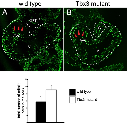
Immunohistochemical analysis of cell proliferation in E9.5 wild type (A) and Tbx3 mutant (B) hearts using antibodies against BrdU (A, B) or phospho-Histone 3 (C). (A, B) Representative sections at the level of the AVC of wild type and mutant hearts are shown. The heart is outlined by a discontinuous white line. The density of BrdU positive cells is higher in the AVC of mutant hearts than in the AVC of wild type hearts (red arrowheads). (C) The number of phospho-Histone3-positive cells in the AVC was counted in consecutive heart sections at the level of the AVC of four wild type embryos with 30?31 somites and three mutant embryos with 30?32 somites. The average number of mitotic cells for the AVC is represented in a histogram. The number of mitotic cells in the AVC is significantly increased (P<0.010) in mutant embryos (90.7±12.7), compared to wild type embryos (58.3±14.5). A, atrium; AVC, atrioventricular canal; OFT, outflow tract; V, ventricle. |
|
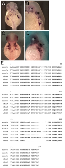
Zebrafish tbx3b and tbx2a are orthologues of mouse Tbx3 and Tbx2. Dorsal (A, C) and lateral views (B, D) of in situ hybridized 48 hpf embryos with tbx3b (A, B) and tbx2a (C, D). Asterisk indicates the fin bud. (E) Comparison of the amino acid sequence of the T-box domain of several Tbx2 subfamily proteins shows that tbx3b and tbx2a exhibit high similarity with other members from this family. EXPRESSION / LABELING:
|

Unillustrated author statements PHENOTYPE:
|

