- Title
-
Sp5l is a mediator of Fgf signals in anteroposterior patterning of the neuroectoderm in zebrafish embryo
- Authors
- Sun, Z., Zhao, J., Zhang, Y., and Meng, A.
- Source
- Full text @ Dev. Dyn.
|
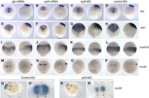
Regulatory effect of sp5l on expression of neuroectodermal genes. Expression sites: fez in the forebrain at the bud stage (A-D), otx1 in the forebrain and midbrain at the 90% epiboly stage (E-H), hoxb1b in the posterior neuroectoderm at the 90% epiboly stage (I-L), and krox20 in the third rhombomere (r3) at the bud stage (M-P) and in r3 and r5 at the 18-somite stage (Q,Q′,R,R′). A-L: Dorsal views with animal pole to the top. M,N: Dorsal views of the hindbrain with anterior to the left. Embryos were injected with 100 pg of gfp mRNA (A,E,I,M), 100 pg of sp5l mRNA (B,F,J,N), 5 ng of sp5l-MO (C,G,K,O,R,R′), or 5 ng control MO (D,H,L,P,Q,Q′). A-P: In each panel on the left is the dorsal view of an embryo with an animal pole to the top and on the right is the lateral view of the same embryo. Q,R: Lateral views with anterior to the left. Q′,R′: Dorsal views showed enlarged hindbrain region in Q and R, respectively. EXPRESSION / LABELING:
|
|
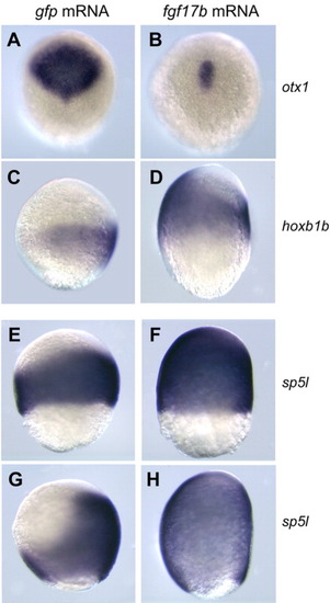
Effect of ectopic Fgf signal on neuroectodermal genes. Embryos were injected with 10 pg of fgf17b mRNA (B,D,F,H) or 100 pg of gfp mRNA (A,C,E,G) at the one-cell stage and examined later by in situ hybridization for expression of otx1 at the bud stage (A,B), hoxb1b at the 90% epiboly stage (C,D), sp5l at the 75% epiboly (E,F), and 90% epiboly (G,H) stages. When injected with fgf17b mRNA, otx1 expression was retained only in the prechordal plate (B), and the expression domains of hoxb1b (D) and sp5l (F,H) expanded ventrally and shifted anteriorly. A,B: Dorsal views with animal pole to the top. C-H: Lateral views with dorsal to the right and animal pole to the top. |
|
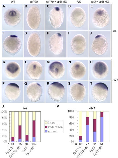
sp5l is required for inhibitory effect of ectopic Fgf signals on anterior neuroectodermal genes. Embryos were injected with 2 pg of fgf17b mRNA (B,G,L,Q), 2 pg of fgf17b mRNA plus 5 ng of sp5l-MO (C,H,M,R), 100 pg of fgf3 mRNA (D,I,N,S), or 100 pg of fgf3 mRNA plus 5 ng of sp5l-MO (E,J,O,T). A,F,K,P: Uninjected wildtype embryos (WT). A-E, K-O: Dorsal views with animal pole to the top. F-J, P-T: Lateral views with animal pole to the top and dorsal to the right. The expression of fez was examined at the bud stage and otx1 at the 90% epiboly stage. Ectopic Fgf signals eliminated or reduced the expression of fez (B,D,G,I) and otx1 (L,N,Q,S). The expression of fez (C,E,H,J) and otx1 (M,O,R,T) could be recovered when endogenous sp5l was simultaneously knocked down with the antisense morpholino. Statistical data were shown in U and V. Embryos were classified into three types according to expression patterns: loss of expression (loss), reduced expression domain (reduction), and normal expression domain (normal). The observed numbers are indicated under each bar. EXPRESSION / LABELING:
|
|
sp5l is required for promoting the effect of ectopic Fgf signals on posterior neuroectodermal genes. Embryos were uninjected (WT) (A,D,G), or injected with 2 pg of fgf17b mRNA (B,E,H) or 2 pg of fgf17b mRNA plus 5 ng of sp5l-MO (C,F,I), and examined by in situ hybridization for the expression of gbx2 at the bud stage (A-C), hoxb1b at the 90% epiboly stage (D-F), and krox20 at the bud stage (G-I). All embryos were shown in lateral views with animal pole to the top and dorsal to the right. Embryos injected with fgf17b mRNA had an elongated axis and showed an anterior shift and ventral expansion of the marker gene expression domains (B,E,H). Coinjection with sp5l-MO inhibited the effect of fgf17b overexpression (C,F,I). Statistical data are summarized in J. The affected embryos were those that showed anterior shift and ventral expansion of the expression domain. Compared to wildtype and fgf17b-injected embryos, some of affected embryos that were coinjected with fgf17 mRNA and sp5l-MO showed weaker expression though the expression domain still shifted anteriorly and expanded ventrally. EXPRESSION / LABELING:
|
|
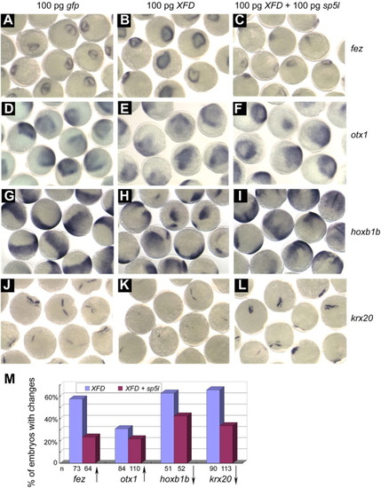
Overexpression of sp5l rescues effects of XFD overexpression in neuroectodermal patterning. Embryos were injected with 100 pg of gfp mRNA (A,D,G,J), 100 pg of XFD mRNA (B,E,H,K), or 100 pg of XFD mRNA plus 100 pg of sp5l mRNA (C,F,I,L), and examined for the expression of fez and krx20 at the bud stage and the expression of otx1 and hoxb1b at the 90% epiboly stage. M: Statistical data indicated percentage of embryos with decreased or increased expression of the marker genes. Upward and downward arrows indicated increase or decrease of expression, respectively. The observed numbers of embryos were indicated below the horizontal axis. |
|
Effect of sp5l on expression of fgf8 and fgf3. Embryos were injected with 5 ng of sp5l-MO (B,E,H,K) or 100 pg of sp5l mRNA (C,F,I,L) at the single cell stage and examined for fgf8 and fgf3 expression by whole-mount in situ hybridization at the end of gastrulation. A-C, G-I: Dorsal views with animal pole to the top. D-F, J,K: These were the same embryos as in A-C and G-I, respectively, but laterally placed with animal pole to the top and dorsal to the right. Note that sp5l knockdown led to the reduction and posterior shift of the presumptive hindbrain domain (hb), and that sp5l overexpression resulted in expansion and anterior shift of this domain. hb, the presumptive hindbrain; fb, forebrain; pm, prechordal mesoderm. EXPRESSION / LABELING:
|


