- Title
-
Expression of cadherin10, a type II classic cadherin gene, in the nervous system of the embryonic zebrafish
- Authors
- Liu, Q., Duff, R.J., Liu, B., Wilson, A.L., Babb-Clendenon, S.G., Francl, J., and Marrs, J.A.
- Source
- Full text @ Gene Expr. Patterns
|
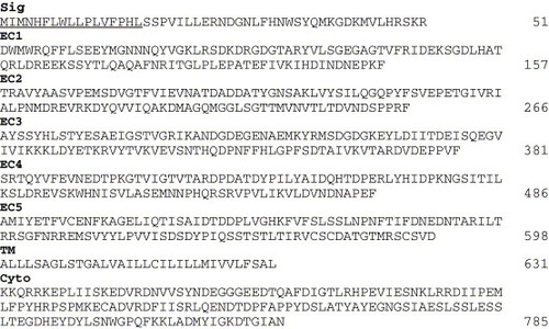
Deduced amino acid sequence of zebrafish Cdh10. The putative hydrophobic signal sequence (Sig) is underlined. Other abbreviations: cyto, cytoplasmic domain; EC1-EC5, extracellular domains 1-5; and TM, transmembrane domain. |
|
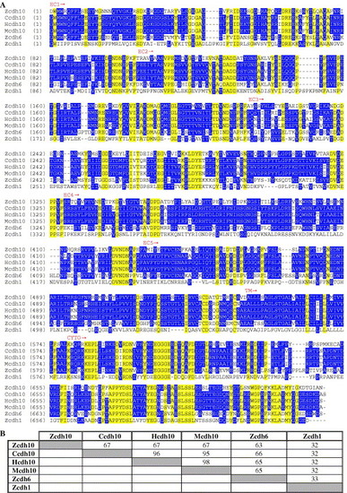
(A) Amino acid sequence comparison between the deduced zebrafish Cdh10 amino acid sequence (Zcdh10), chicken Cdh10 (Ccdh10), human Cdh10 (Hcdh10), mouse Cdh10 (Mcdh10), zebrafish Cdh6 (Zcdh6), and zebrafish Cdh1 (Zcdh1). Comparisons were between published sequences from the EC1 to the end of the coding sequences. Sequences highlighted by yellow boxes indicate residues that are common to all six sequences, and sequences highlighted by blue boxes indicate amino acids that are identical to at least half of the sequences. (B) Sequence identity percentages for pairwise comparisons between all six sequences shown in the alignment. Diagonal shaded boxes indicate sequence comparisons between the same sequences, and therefore, represent 100% identity. Sequence comparisons were performed using Align X (InforMax Inc., North Bethesda, MD). Abbreviations the same as in Fig. 1. |
|
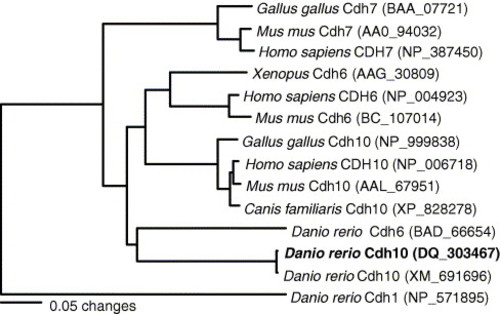
Phylogram resulting from neighbor-joining distance analysis of EC1 through the carboxy-terminal protein sequence alignment. The tree was rooted with the zebrafish Cdh1 amino acid sequence. GenBank accession numbers follow the sample names. The sequence generated as part of this study is shown in bold. |
|
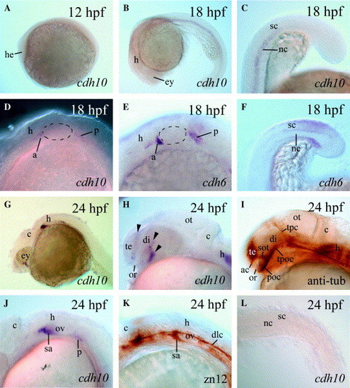
cdh10 expression in 12?24 hpf zebrafish embryos. All panels show lateral views of whole mount embryos labeled with cdh10 cRNA (A?D, G, H, J, and L), cdh6 cRNA (E and F), anti-acetylated tubulin (anti-tub, I) or zn12 (K) antibodies. Panels (D), (E), (H), (I), (J), and (K) are higher magnifications of the head region (anterior to the left and dorsal up), while (C), (F), and (L) are higher magnifications of the posterior trunk and tail region (due to the bend between the trunk and tail, these two regions have different orientations: for (C) and (F), anterior down and dorsal to the left for the posterior trunk region, while anterior to the left and dorsal up for the tail region; for (L), anterior to the left and dorsal up for the trunk region, while anterior to the left upper corner and dorsal to the right upper corner). The otic placode is outlined with dashed lines. Each of three arrowheads in (H) points to a cdh10 expression strip in the forebrain. Abbreviations: a, anterior lateral line placode area; ac, anterior commissure; c, cerebellum; di, diencephalon; dlc, dorsal longitudinal tract; ey, eye; he, head region; h, hindbrain; nc, notochord; or, optic recess; ot, optic tectum; ov, otic vesicle; p, posterolateral line placode/ganglion; poc, postoptic commissure; sa, statoacoustic ganglion; sc, spinal cord; sot, supraoptic tract; sp, spinal cord; te, telencephalon; tpc, tract of posterior commissure; and tpoc, tract of postoptic commissure. EXPRESSION / LABELING:
|
|
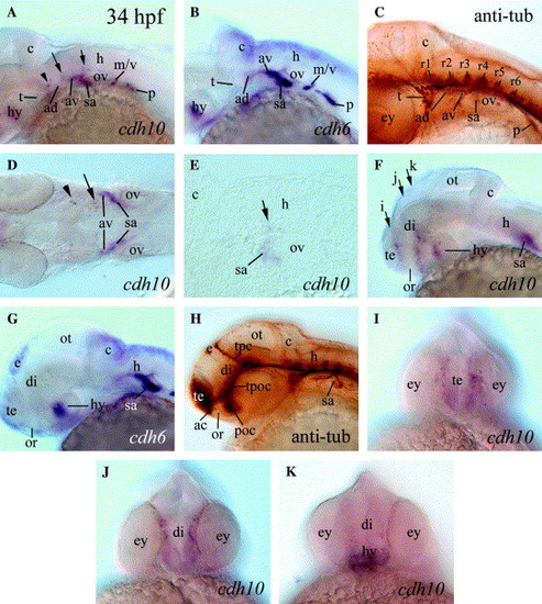
cdh10 expression in 34 hpf embryos (A, D-F, and I-K) compared to cdh6 expression (B and G) and anti-acetylated tubulin staining (C and H). Panels (A-C and F-H) are lateral views of whole mount embryo heads with anterior to the left and dorsal up. (D) A dorsal view (anterior to the left) of a deyolked embryo. (E) A parasagittal section of the hindbrain (anterior to the left and dorsal up). (I)-(K) Frontal views (dorsal up) focusing on planes indicated by three correspondingly labeled arrows in (F). The arrowhead, long and short arrows in (A), (D), and (E) point to the same cdh10 expressing cell clusters. Abbreviations: ad, anterodorsal lateral line ganglion; av, anteroventral lateral line ganglion; hy, hypothalamus; m/v, medial lateral line/vagus ganglion, r1-r6, rhombomeres 1-6; and t, trigeminal ganglion. The remaining abbreviations are the same as in Fig. 4. EXPRESSION / LABELING:
|
|
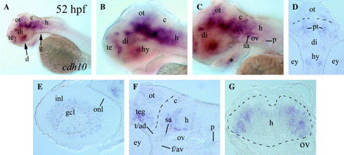
cdh10 expression in 52 hpf embryos. Panels (A?C) are lateral views (anterior to the left and dorsal up) of whole mount embryos. Labeled arrows in (A) indicate levels of cross sections in respective panels. Panels (B) and (C) are higher magnifications of the head region of the embryo in (A), focusing on cdh10 expression domains in more medial and lateral regions, respectively. Panels (E) and (F) are parasagittal sections of the eye and the posterior half of the brain, respectively with anterior to the left and dorsal up. The asterisk in (C) indicates cdh10 expression in the first pharyngeal arch medial to the lower jaw. The dashed line in (D) indicates the border between the thalamus and optic tectum. The dashed line in (F) indicate the boundary between the mid- and hindbrains. The hindbrain border in (G) is outlined by the dashed line. Abbreviations: f/av, facial and anteroventral lateral line ganglion; gcl, retinal ganglion cell layer; inl, inner nuclear layer; onl, outer nuclear layer; pt, pretectal area; teg, tegmentum; t/ad, trigeminal; and anterodorsal lateral line ganglion. The remaining abbreviations are the same as in the previous figures. EXPRESSION / LABELING:
|

Unillustrated author statements |
Reprinted from Gene expression patterns : GEP, 74(6), Liu, Q., Duff, R.J., Liu, B., Wilson, A.L., Babb-Clendenon, S.G., Francl, J., and Marrs, J.A., Expression of cadherin10, a type II classic cadherin gene, in the nervous system of the embryonic zebrafish, 1016-1025, Copyright (2006) with permission from Elsevier. Full text @ Gene Expr. Patterns

