- Title
-
Expression analysis of jagged genes in zebrafish embryos
- Authors
- Zecchin, E., Conigliaro, A., Tiso, N., Argenton, F., and Bortolussi, M.
- Source
- Full text @ Dev. Dyn.
|
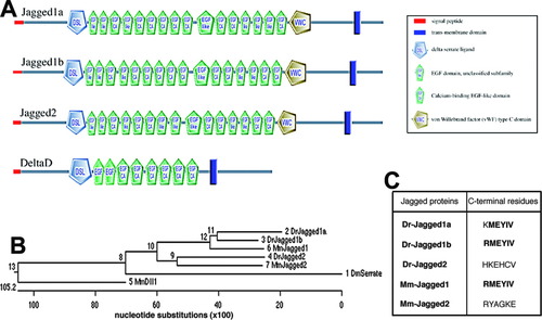
Domain structure and phylogenetic relationships of zebrafish Jagged proteins. A: Schematic diagram of the motif/domain structure of zebrafish Jagged1a, Jagged1b, Jagged2, and DeltaD, obtained with the SMART program (http://smart.embl-heidelberg.de). The cysteine-rich domain encompasses the von Willebrand factor type C-like domain. B: Dendrogram of zebrafish (Danio rerio, Dr) Jagged1a, Jagged1b, and Jagged2; mouse (Mus musculus, Mm) Jagged1, Jagged2, and Dll1 (Delta-like1); and Drosophila (Drosophila melanogaster, Dm) Serrate, obtained with the MegAlign program that uses the Clustal W algorithm (Thompson et al., [1994]). C: C-terminal hexapeptides of zebrafish (Dr) and mouse (Mm) Jagged proteins. The hexapeptide of Jagged1b is identical, whereas that of Jagged1a differs by one amino acid, from the sequence of mouse Jagged1, shown to represent a functional PDZ-ligand (Hock et al., [1998]; Ascano et al., [2003]). The conserved amino acids in the three hexapeptides are in bold characters. The hexapeptides of zebrafish and mouse Jagged2 are not conserved and neither resemble a PDZ-ligand (Hock et al., [1998]; Ascano et al., [2003]). |
|
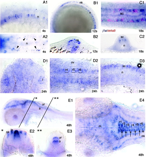
jagged1a expression pattern. in situ hybridization of embryos at different developmental stages performed with the jagged1a probe. A: At the four-somite stage (4s), jagged1a is expressed in the notochord (A1) and in scattered cells (arrowheads) in the intermediate neural plate (A2). B: At the 12-somite stage (B1, B2) jagged1a is expressed in the notochord and in cells located in the intermediate neural keel. C: At the 18-somite stage (C1, C2) the notochord is no longer labeled, while in the spinal cord jagged1a is expressed in cells located in a mediolateral position where some cells expressing deltaB are also observed (C1). D: At 24 hpf jagged1a is expressed in the lens (D1), the rhombomere borders (D2), and the spinal cord (D3). E: At 48 hpf in the central nervous system (E1-E4), jagged1a-expressing cells occur in the midbrain, hindbrain, and spinal cord. In the hindbrain (E1, E2, E4), the labeled cells are located dorsally at the anterior and posterior rhombomere borders and in bilateral inner and outer strings of cell clusters. In the caudal hindbrain and spinal cord (E1 and E3), the jagged1a-expressing cells are situated medially in bilateral upper and lower stripes. In D2 and E4, the position of the rhombomeres has been determined on the basis of the location of the otic vesicles. The arrows in E4 indicate the boundaries between the rhombomeres. Views are lateral (A1, B1, D3, E1) or dorsal (A2, C1, D1, D2, E4); transverse sections are the level of the trunk (B2, C2), the rhombomeres (E2), and caudal hindbrain (E3). Anterior is to the left in all lateral and dorsal views except for D1, in which it is to the top. is, inner string of jagged1a-expressing cell clusters; l, lens; lr, lower row of jagged1a-expressing cells; m, midbrain; n, notochord; nk, neural keel; os, outer string of jagged1a-expressing cell clusters; ov, otic vesicle; r1 to r6, rhombomeres 1 to 6; sc, spinal cord; ur, upper row of jagged1a-expressing cells. EXPRESSION / LABELING:
|
|
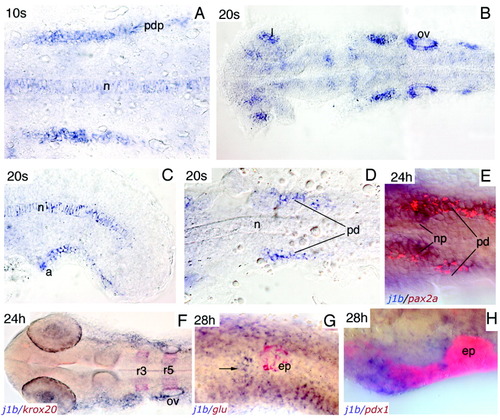
jagged1b expression pattern. In situ hybridization of embryos at different developmental stages performed with the jagged1b probe. A: At the 10-somite stage (10s), jagged1b is expressed in the notochord and the pronephros primordia. B-D: At the 20-somite stage, jagged1b-expressing cells are detected mainly in the lens, otic vesicles, and two bilateral groups of cells situated anteriorly and posteriorly to the latter (B), in the caudal part of the notochord (C), and in the rostral part of the pronephric ducts (D). C: At the 20-somite stage, jagged1b transcripts are detected in a group of cells located ventrally and posteriorly to the anus. E: At 24 hours postfertilization (hpf), jagged1b and pax2a are coexpressed in cells of the rostral end of the pronephric duct and lateral part of the nephron primordia, but only jagged1b is expressed in the medial cells of the nephron primordia. F: At the same stage, jagged1b transcripts are also expressed in the region extending from the otic vesicle to the eye. G,H: At 28 hpf, the jagged1b (arrow) expression domain does not overlap with that of glucagon in the pancreatic islet (G), but is in part overlapped with the pdx1 expression domain (H). Views are dorsal (A,B,F), lateral (C,H), or ventral (D,E,G). Anterior is to the left in all pictures. a, anus; ep, endocrine pancreas; l, lens; n, notochord; np, nephron primordia; ov, otic vesicle; pd, pronephric duct; pdp, pronephric duct primordium; r3, rhombomere 3; r5, rhombomere 5. EXPRESSION / LABELING:
|
|
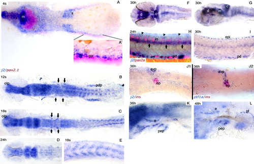
jagged2 expression pattern. In situ hybridization of embryos at different developmental stages performed with the jagged2 probe. A: At the four-somite stage (4s), jagged2 is expressed in the mesoderm as shown by colabeling with the pax2a probe (see inset A′. B,C: At the 12- and 18-somite stages, jagged2 is strongly expressed throughout the brain, in the developing somites (shown at a higher magnification in E), the pronephric duct primordia, and in two clusters of cells (arrows) in the otic placode (B) and vesicle (C). D-G: At 24 hours postfertilization (hpf, D) and 30 hpf (F,G), jagged2 is expressed in the diencephalon, midbrain, and cerebellum, and with regard to the 30-hpf stage, in the ventromedial part of the rhombomeres as well. H: At 24 hpf, jagged2 is expressed strongly and diffusely in the ventral part of the spinal cord (arrows) and faintly in the dorsal part (arrowheads) but not in interneurons located in between detected by pax2a labeling. Moreover, jagged2 is also expressed in scattered cells of the pronephric duct, in which some pax2a-expressing cells are also observed. I: At 30 hpf, the jagged2 expression pattern is identical to that observed at 24 hpf, but the picture evidences the expression in the epidermis, which is out of focus in H. J1,J2: jagged2 labeling is observed in the anteroventral pancreatic anlage that expresses ptf1a and grows to engulf the endocrine pancreatic islet identified by insulin expression. K,L: At 36 hpf (K) and 48 hpf (L), jagged2 is expressed in the ear, pharyngeal pouches, and pectoral fin. Views are dorsal (A-D,F), lateral (E,G-I,K,L), or ventral (J1,J2). Anterior is to the left in all pictures. avp, anteroventral pancreas; c, cerebellum; d, diencephalon; e, ear; ep, endocrine pancreas: epi, epidermis; in, interneurons; m, midbrain; olp, olfactory placode; pep, pharyngeal endodermal pouches; pd, pronephric duct; pdp, pronephric duct primordium; pf, pectoral fin; pp, prospective pronephros; s, somites. EXPRESSION / LABELING:
|

Unillustrated author statements EXPRESSION / LABELING:
|


