- Title
-
Fgf signaling is required for zebrafish tooth development
- Authors
- Jackman, W.R., Draper, B.W., and Stock, D.W.
- Source
- Full text @ Dev. Biol.
|
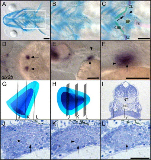
Zebrafish teeth are located deep in the posterior, ventral portion of the pharynx, but can be visualized by several methods. (A?C) Ventral views of 5 day larvae stained with alcian green to label cartilages and highlight mineralized teeth, anterior to the left. (A) A broad, ventral view of the head shows the area where teeth develop (box). (B and C) Unlabeled and labeled views of the posterior pharynx. The earliest-formed teeth (arrow) are by this time attached to the 5th ceratobranchial cartilage (CB5), while the third-formed teeth (arrowhead) have just begun to mineralize and are not yet attached. The second tooth pair is out of the plane of focus. Elements of the pectoral girdle flank the tooth-forming region (scapulocoracoid, SC; cleithrum, CL), the esophagus lies caudally (ES), and the keratinized bite pad (carpstone), against which the teeth ultimately bite, is present in the midline on the dorsal surface of the pharynx (BP). (D?F) Expression of dlx2b at 56 h reveals the location and orientation of the first pair of tooth germs, before they have begun to mineralize. Anterior to the left. (D) Dorsal view focused through the hindbrain, arrows indicate the tooth germs. (E) Lateral view shows a tooth germ (arrow) just ventral and rostral to the first myotome (arrowhead). (F) Close-up of (E) reveals that the dental epithelium (arrow) surrounds the dental mesenchyme except rostrally (arrowhead). (G and H) Diagrams of 56 h tooth germs with the dental mesenchyme colored dark blue, and the dental mesenchyme a lighter shade. (G) Tooth germ viewed from dorsal, mimicking the orientation of the left-side tooth germ in (D). Transverse planes of section are indicated (J?L). (H) Tooth germ viewed laterally as in (E) and (F). (I) Transverse section of a 56-h larvae at the level of the developing teeth to indicate the location of the left-side tooth germ in this plane of section (box; NT, neural tube; NC, notochord; Y, yolk; up is dorsal). (J?L) Three typical shapes of a 56-h tooth germ in transverse section. (J) At the rostral end, a group of dental mesenchyme cells (dark blue, arrowhead) is flanked medially by a crescent of dental epithelium (light blue, arrow). (K) In the center of the tooth germ, the epithelium (arrow) completely surrounds a core of mesenchyme (arrowhead). (L) Caudally, the section does not pass through the mesenchyme at all, and only dental epithelial cells are seen (arrow). Scale bars = 100 μm. |
|
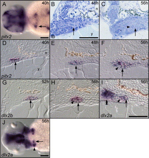
pitx2 expression is the earliest indicator of zebrafish tooth development, and Dlx2 orthologs the earliest specific tooth markers we identified. (A) At 36 h (40 h shown), pitx2 is expressed broadly in the pharyngeal epithelium, with the strongest expression prefiguring the first tooth germs (arrow, dorsal view, anterior to the left). (B) At 48 h, the dental epithelium has begun to thicken and undergo morphogenesis (arrow). (B?I) Transverse sections oriented as in Fig. 2I. (C) By 56 h, the tooth germ has undergone morphogenesis with the dental epithelium (arrow) surrounding a core of mesenchyme (arrowhead). (D) At 40 h, pitx2 is expressed in the pharyngeal epithelium (arrow), but no sign of dental morphogenesis is yet visible. (E) At the beginning of dental morphogenesis at 48 h, pitx2 expression is maintained in the pharyngeal epithelium (arrow). (F) At 56 h, pitx2 expression continues in the pharyngeal epithelium, including the dorsal and medial dental epithelium (arrow) adjacent to the non-expressing mesenchyme (arrowhead). (G) dlx2b expression is first detectable in the dental epithelium at 48 h (arrow, 52 h shown), and continues to be expressed in the epithelium (arrow) and mesenchyme (arrowhead) at 56 h (H). (I and J) dlx2a is expressed in the dental epithelium (arrows) and mesenchyme (arrowheads) at 56 h, but in contrast to dlx2b, is also expressed in lateral arch mesenchyme (double-arrowhead). Labels: n = notochord, y = yolk. Scale bars = 100 μm. |
|
fgf3 and fgf4 are expressed in the dental epithelium and lhx6 and lhx7 in the dental mesenchyme, but we did not detect fgf8 or pax9 expression in zebrafish tooth germs. (A) fgf8 expression is detectable in pharyngeal pouches until at least 44 h (arrowhead), but we do not detect it in tooth germs at any stage examined (arrow). (B) fgf4 is expressed in the dental epithelium at 48 h (arrow), as well as in lateral pharyngeal endoderm (arrowhead). (C) Transverse section at 56 h shows fgf4 expression localized to a subset of the dental epithelium (arrow). (D) fgf3 is expressed in a very similar pattern to fgf4 in the dental epithelium (arrow) and in the pharyngeal pouches (arrowhead), but is not detectable in the dental epithelium until 52 h. (E) At 56 h, fgf3 is expressed in what appears to be an identical subset of the dental epithelium as fgf4 (arrow). (F) lhx6 expression is detectable in lateral pharyngeal mesenchyme (arrowhead) and dental mesenchyme (arrow) at 48 h. (G) This lateral (arrowhead) and dental mesenchyme (arrow) expression is maintained at 56 h. (H and I) In the region of tooth formation at 56 h, lhx7 expression is localized to the dental mesenchyme (arrows). (J) pax9 expression is visible laterally in the pharyngeal arches at 48 h (arrowhead) but not in the tooth-forming region (arrow). (K and L) At 56 h, pax9 continues to be visible in the non-dental pharyngeal epithelium (double-arrows) and in lateral pharyngeal mesenchyme (arrowheads), but pax9 expression is undetectable in the tooth germs (arrows). (A, B, D, F, H, and J) Dorsal views, anterior to the left. (C, E, G, I, K, and L) transverse sections as in Fig. 2I. Scale bars = 100 μM. |
|
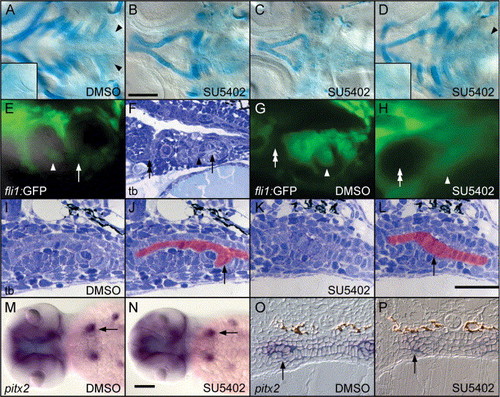
SU5402 inhibits zebrafish pharyngeal tooth morphogenesis. (A?D) Ventral views of 82-h cartilage-stained specimens centered on the posterior pharynx and focused at the level of the teeth and ceratobranchial cartilages, anterior to the left. (A) Control larvae treated from 32 to 82 h with 0.5% DMSO develop normal pharyngeal cartilages and teeth (arrowheads and inset). (B) Larvae treated from 32 to 82 h with 25 μM SU5402 exhibit pharyngeal cartilage reduction, and teeth are absent. However, occasionally, specimens are seen in these treatments with more severe cartilage reductions (C), or with more normal cartilages and teeth present (arrowhead and inset, D). (E) A fluorescent/bright-field double image of a 78-h tooth germ in the fli1:GFP transgenic line. Non-GFP-expressing dental epithelium (mineralized portion of tooth indicated with arrow) surrounds GFP-expressing dental mesenchyme (arrowhead). (F) A toluidine blue-stained sagittal section at 72 h showing the dental mesenchyme (arrowhead), mineralized tooth tip (arrow), and the location of the 6th pharyngeal pouch (double-arrow). (G) In a control larva treated with 0.5% DMSO from 32 to 78 h, the GFP-expressing dental mesenchyme (arrowhead) surrounded by non-expressing epithelium is located caudad to the 6th pouch (double-arrow). (H) In 32?78 h SU5402-treated individuals, the 6th pharyngeal pouch has adopted a rounded morphology (double-arrow), and no gap in GFP expression is detectable in the region where the tooth would normally form (arrowhead). (I) Toluidine blue stained transverse section of the tooth germ after DMSO exposure from 32 to 56 h. The pharyngeal epithelium is colored in red in (J), with the curved dental epithelium visible (arrow). (K and L) After 32?56 h SU5402 treatment, morphogenesis of the dental epithelium is no longer apparent, although this epithelium may be slightly thickened (arrow). (M and N) Expression of pitx2 in the tooth germs is identical between 32?56 h DMSO-treated controls (arrow, M) and 32?56 h SU5402-treated individuals (arrow, N). Dorsal views, anterior to the left. (O) Likewise, transverse sections reveal normal pitx2 expression and dental epithelial morphogenesis at 56 h in DMSO control embryos (arrow), while in 32-56 h SU5402-treated specimens (P), pitx2 expression remains in the pharyngeal epithelium but no epithelial morphogenesis is visible (arrow). Scale bars = 100 μM. EXPRESSION / LABELING:
|
|
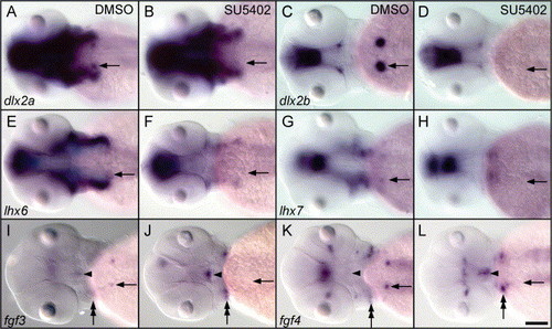
Dlx, Fgf, and Lhx tooth-related gene expression is inhibited by SU5402 treatment. Dorsal views of 56 h in situ hybridizations, focused at the level of the pharynx, anterior to the left. Embryos were treated with 0.5% DMSO (columns A and C) or DMSO+ 25 μM SU5402 (cols. B and D) from 32 to 56 h. The location of the left side tooth germ is indicated by an arrow in all panels. Tooth germ expression of dlx2a, dlx2b, lhx6, lhx7, fgf3, and fgf4 is present in DMSO controls, but absent in SU5402-treated specimens. (I?L) fgf3 and fgf4 expression in the ventral diencephalon (arrowheads) and in the anterior pharyngeal pouches (double arrows) appears stronger in Su5402-treated specimens (J and L). Scale bar = 100 μM. |
|
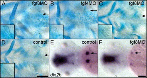
Teeth develop relatively normally after morpholino antisense inhibition of fgf3 and fgf8, but subtle tooth effects are observed after fgf4 inhibition. (A?D) Ventral view of the pharyngeal region of morpholino (MO) injected, cartilage-stained specimens at 5 days with magnified teeth shown in inset. (A) Ceratobranchial cartilages were often completely missing after fgf3 MO injection, but teeth appeared normal (arrow). (B) After fgf4 MO injection, ceratobranchial cartilages were also reduced, and teeth, although always present, were often misshapen (arrow). (C) fgf8 morpholino injected fish display severe cartilage reductions, but teeth always developed normally (arrow). (D) Injection control displaying normal cartilages and teeth (arrow). (E and F) Dorsal views of control and fgf8 MO injected fish at 56 h. dlx2b expression in tooth germs (arrow, E) was variably affected: sometimes normal, sometimes missing, and sometimes reduced (arrow, F). Anterior is to the left in all panels. Scale bars = 100 μM. EXPRESSION / LABELING:
|
Reprinted from Developmental Biology, 274(1), Jackman, W.R., Draper, B.W., and Stock, D.W., Fgf signaling is required for zebrafish tooth development, 139-157, Copyright (2004) with permission from Elsevier. Full text @ Dev. Biol.

