- Title
-
Development of a rod photoreceptor mosaic revealed in transgenic zebrafish
- Authors
- Fadool, J.M.
- Source
- Full text @ Dev. Biol.
|
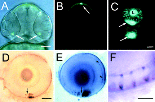
Comparison of EGFP and rhodopsin expression in transgenic zebrafish. EGFP expression in photoreceptor cells (arrows) of the ventral retina (A) and the pineal (B) of transgenic zebrafish larva observed at 60 hpf. (C) At 84 hpf, EGFP fluorescence could be observed in the ventral patch (arrows) of the retinas and in cells sporadically positioned across the retina. Apparent fluorescence by the lens (L) is due to photoreceptor cell expression. In situ hybridization of EGFP (D) and rhodopsin (E) demonstrate a similar pattern of labeling of cells in the outer nuclear layer (ONL) of the ventral retina and on opposite sides of the choroid fissure (arrow). Sporadic labeling of cells in the ONL is also observed with the opsin probe (arrowheads). (F) Double labeling demonstrated colocalization of the EGFP transcript (brown) and rhodopsin transcript (purple) to sporadically spaced cells located in the outer nuclear layer of larval fish. Bar, 50 mm in (A?C); 25 mm in (D, E); 10 mm in (E). |
|
Confocal analysis of EGFP expression of rod photoreceptors. Whole retinas maintained in saline were examined by confocal microscopy. (A) Low magnification image of the whole explant demonstrated robust expression of EGFP across the entire retina. (B) A higher magnification composite image of scans taken near the retinal margin. Cells with a characteristic rod morphology highlighted by small terminals (te), round soma, long thin inner segments (is), and rod-shaped outer segments (os) were observed. Images taken through the ganglion cell layer and tangential to the surface of the eye reveal a distinct organization of the rod photoreceptors at the level of the inner segments (C), cell bodies (D), and terminals (E). Note that, in each image, the rod structures are positioned in regularly spaced rows and a seam where two planes of the mosaic merge. Bar, 5 mm in (B); 20 mm in (C?E). |
|
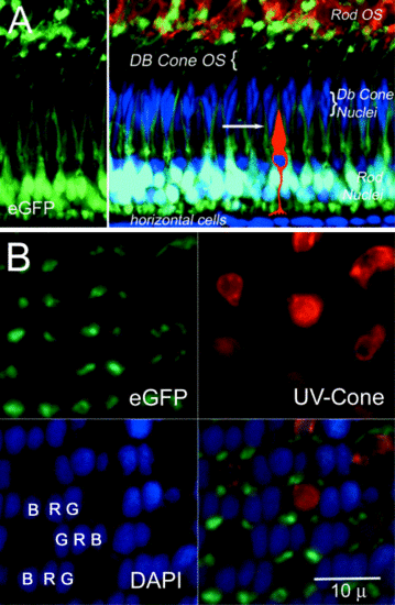
Comparison of the rod mosaic to cone mosaic. (A) Transverse section through the adult retina demonstrating EGFP fluorescence in rod photoreceptors (left panel in green) and counterstaining with DAPI (blue) to reveal the tiering of the cone nuclei (left merged image). The rod cell bodies appear white due to double labeling for EGFP and DAPI. The rod outer segments are also immunolabeled for opsin. The position of the short single cone is diagrammatically represented and the plane of section in (B) is indicated by the arrow. (B) EGFP fluorescence of the rods, immunolabeling of the UV sensitive opsin, DAPI labeling of the cone nuclei in a tangential section of the adult retina and the merged image. Note the regular arrangement of the rod myoids around the outer segment of the UV-cone outer segments. (B, blue cone; G, green cone; R, red cone). |
|
Rod differentiation at the retinal margin. (A?C) EGFP expression, immunolabeling for rhodopsin (Rod), and the merged image of an oblique section through the retinal margin reveal regularly spaced clusters of rods (numbered 1?7). (D?F) Merged images of immunolabeling (red) for rhodopsin (D), UV opsin (E), and red opsin (F) with EGFP expression (green) and DAPI nuclear staining (blue) of serial sections taken parallel to the retinal margin. Note the positions of the clustered rod outer segments at the positions of the immature UV cone outer segments (arrows). |
|
Rod photoreceptor mosaic development in the larval retina. (A) Merged stack of low magnification confocal images of EGFP expression in the eye from a 10-dpf larval fish. The fluorescence appears scattered with regions of high density and obvious gaps in the distribution of rods. (B) At a higher magnification, the regular arrangement of parallel row of rods is indicated (arrows). (C) Histological section to demonstrate the regular spacing of the short single cones and the double cones in the larval retina. (D) Confocal image of EGFP fluorescence in larva 21 dpf. Note the regular arrangement of rows and the numerous telodendria extending from the rod terminals (arrowheads) Bars, 50 μm (A), 10 μm (B), 7 μm (C, D). |
Reprinted from Developmental Biology, 258(2), Fadool, J.M., Development of a rod photoreceptor mosaic revealed in transgenic zebrafish, 277-290, Copyright (2003) with permission from Elsevier. Full text @ Dev. Biol.


