- Title
-
The zebrafish unplugged gene controls motor axon pathway selection
- Authors
- Zhang, J. and Granato, M.
- Source
- Full text @ Development
|
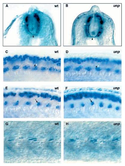
Spinal axonal trajectories, motor neuron specification, and muscle pioneer development appear unaffected in unplugged mutant embryos. (A,B) Cross-section of 27 hpf embryos stained with anti-acetylated tubulin. In the wild-type (A) and the mutant embryo (B), a commissural primary ascending (CoPA) soma (white arrowhead) extends an axon ventrally to the midline (black arrowhead) and across the midline. Staining with islet-1 antisense probe (C,D) or islet-2 antisense probe (E,F) of a 27 hpf wild-type (C,E) and an unplugged mutant embryo (D,F), lateral views. Black and white arrowheads point to isl-1 positive cells (C,D) in the ventral spinal cord. The position of these two cells is consistent with the position of RoP and MiP neurons, respectively. Arrows in E and F point to isl-2 positive cells in the ventral spinal cord, consistent with the position of CaP neurons. In about 50% of the wild-type and unplugged hemisegments, a second motor neuron, VaP, is stained by islet-2 (Appel et al., 1995; Eisen et al., 1990). RoP, MiP and CaP motor neurons were identified by their dorsoventral and rostrocaudal position within the spinal cord, their position relatively to the somite boundary (not in focus) and their soma size (see Materials and Methods for details). Lateral view of a 27 hpf wild-type (G) and an unplugged mutant embryo (H) stained with 4D9, which recognizes the nuclear Engrailed epitope in muscle pioneer cells. About 2-6 muscle pioneer cells are present per hemisegment, at the level of the horizontal myoseptum (Hatta et al., 1991). The number and position of 4D9 positive muscle pioneer cells in unplugged mutant embryos is indistinguishable from those in wild-type embryos. |

