
|
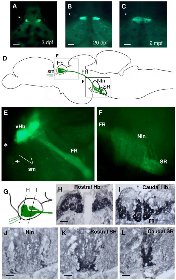
Fig. 4 Spon1b:GFP expression in the habenular nuclei and their projections.
A-C. Dorsal view of live zebrafish showing migration of ventral habenular nuclei from lateral to medial position: 3 dpf (B), 20 dpf (C), and 2 mpf (D). Asterisk indicates the position of the eye. D. Schematic of DCP in adult zebrafish showing areas of spon1b:GFP expression (E and F) and depicting the habenula (Hb), its afferent projections within the stria medullaris (sm), its efferent projection: fasciculus retroflexus (FR), and target nuclei: interpeduncular nucleus (NIn), and superior raphe (SR). E-F. Para-sagittal cut through fresh-frozen adult Tg(spon1b-GFP) brain. GFP fluorescence highlights all DCP structures, including vHb nuclei, FR and sm (E). Also, cells of the bed nucleus of the stria medullaris (BNSM, arrow in E), and projections to NIn and SR in F. Rostral to the left. G. Schematics of the relative shape and position of the spon1b-positive nuclei of the Hb, as shown in H-I. H-L. Coronal sections immunostained for GFP: rostral (H) and caudal (I) Hb, NIn (J), and rostral (K) and caudal (L) SR. Scale bars: A-B: 100 μm; D: 25 μm; H-L: 50 μm.