
Image
|
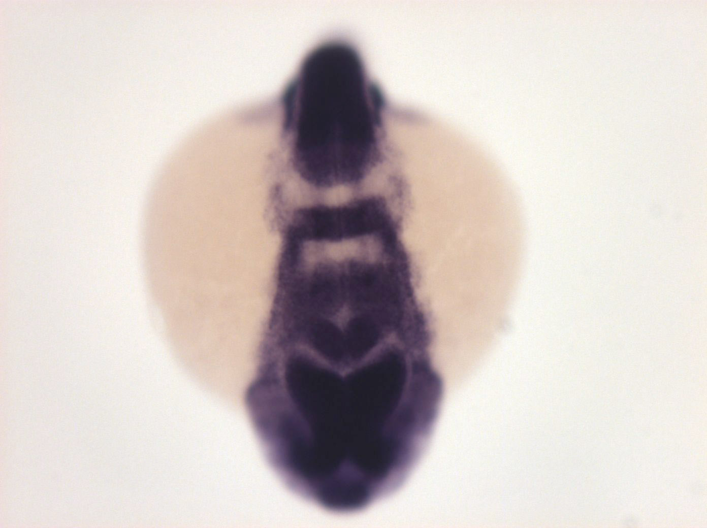
Figure Caption
Fig. 4 anterior forebrain, midbrain, anterior hindbrain, ∅ in rhombomere 2, expressed in rhombomere 3, 5 and 6, ∅ in rhombomere 4, spinal chord, restricted to posterior somites, lateral line
Orientation
| Preparation | Image Form | View | Direction |
| whole-mount | still | frontal | dorsal to top |
Figure Data