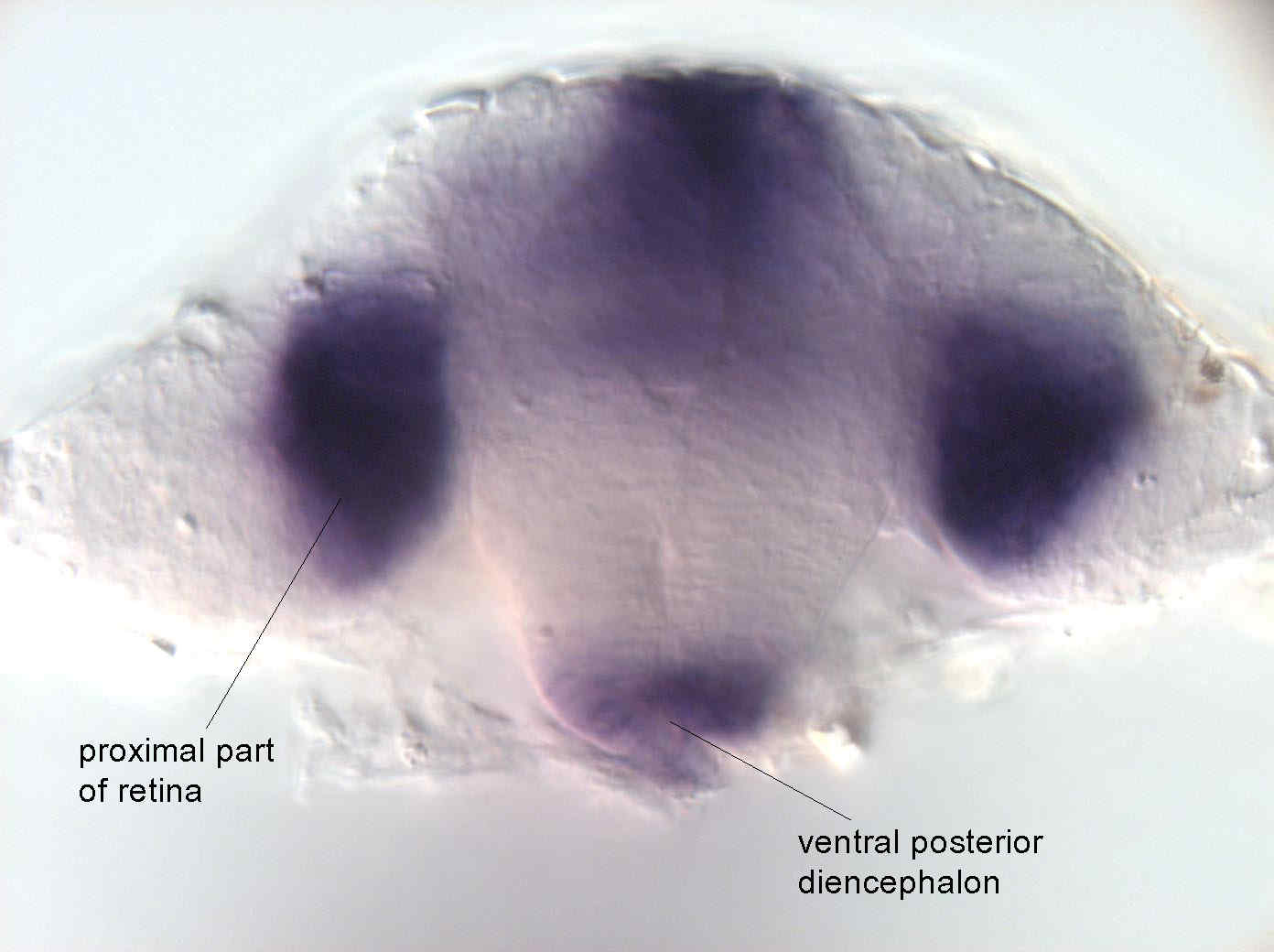
otical cross section of the head at the 20 somite stage at the level of the eye.
Fig. 5 At 20 somite stage, expression increases in the proximal part of retina and the ventral diencephalon. Strong staining is observed in telencephalon, midbrain-hindbrain boundary, optic stalk and the dorsal diencephalon. Moreover, staining in now observed in ventral anterior mesencephalon. In the ear, fgf8 is expressed in the anterior most as well as the dorsal-posterior cells of the otic capsule. Expression is now observed in hyoid arch. Posteriorly, staining is observed in caudal somites as well as in dorsal and caudal fin fold. At 30 hrs expression is also observed in the nose.
| Preparation | Image Form | View | Direction |
| whole-mount | still | transverse | dorsal to top |

