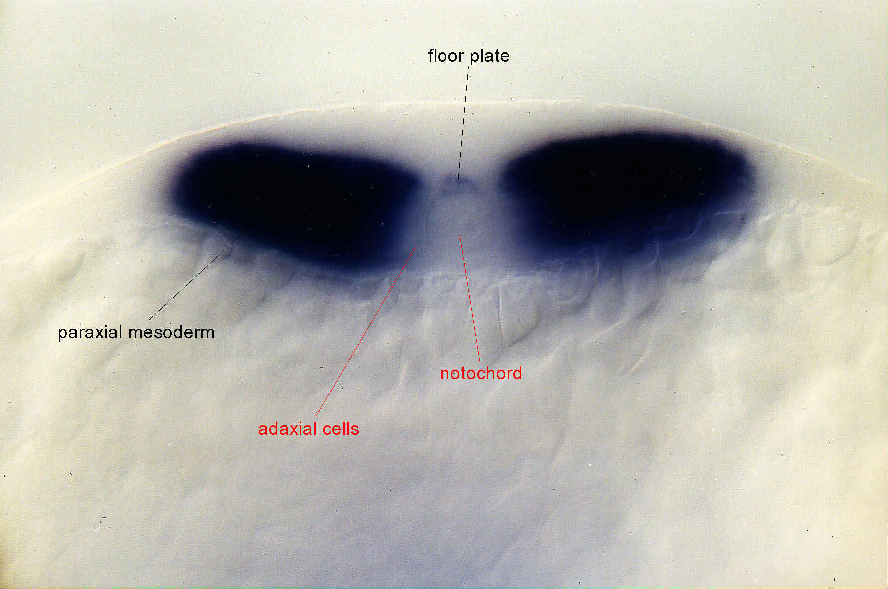
optical cross section at the level of posterior somites at the 6 somite stage.
Fig. 3 The strong stripe in presumptive hindbrain progressively restrict to rhombomere 4 and the ventral part of rhombomere 2. Heart primordia appear labeled by the beginning of somitogenesis. At the 2/3 somite stage, the floor plate, posterior and lateral neural plate appear labeled as well as the caudal most axial tissues. In cross sections, cells located underneath the posterior notochord (endoderm?) are labeled. In hindbrain region, expression stay in the whole rhombomere 4 but is restricted to the ventral most part of rhombomere 2. Around the 3/4 somite stage, anterior expression start to encompass the midbrain-hindbrain boundary. In the paraxial mesoderm, Fgf8 expression is observed in newly formed somites. Anteriorly, after the 2 somite stage, strong labelling is observed in the telencephalon. At the 5 somite stage, expression is strong in the telencephalon, the midbrain-hindbrain boundary, in posterior rhombomere 2 (or anterior part of the cerebellum (?)), ventral part of rhombomere 2. Expression is observed in dorsal and ventral but not the central part of Rhombomere 4. Expression is still observed in heart primordia. In truncal region Fgf8 expression is observed in paraxial somitic mesoderm but not in the adaxial cells. Staining is maintained in the floor plate. By the 8 somite stage, expression has disappeared from the rhombencephalon but persists at the midbrain-hindbrain boundary.
| Preparation | Image Form | View | Direction |
| whole-mount | still | transverse | dorsal to top |

