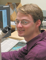
Person
Megason, Sean
|

|
Biography and Research Interest
Sean Megason received a B.S. in Molecular Biology from the University of Texas at Austin in 1996 and a Ph.D. in Molecular Cell Biology at Harvard University in 2001. He conducted post-doctoral research at the California Institute of Technology. Sean joined the Department of Systems Biology at Harvard as an Assistant Professor in April 2008.
Research Summary
Embryonic development is the execution of a program encoded in the genome. I wish to understand how this genomic program is executed to create an embryo in the same way that a computer scientist understands the execution of a computer program. The computer scientist has a significant advantage in understanding how a computer program works because the code for a program is transparent: it can be easily read, browsed, altered, and recompiled in its entirety using a software editor. This of course is not the case for biologists because their object of interest, the embryo, is largely a black box. Research in the Megason lab is focused on using two technologies we developed called in toto imaging and flip traps to bring the same level of transparency and quantitation to the code of life. The ultimate goal of in toto imaging is to ?upload? the embryo into a digital recreation that can be quantitatively and comprehensively studied with same transparency and facility as a complex computer program.
Our approach to doing systems biology in embryos is heavily based on imaging because of its unique ability to capture quantitative data at single cell resolution in living, functioning embryos. We use zebrafish because of their unique suitability for imaging, genetics, and genomics. We are developing a technology called ?in toto imaging? that seeks to track all the cells in a developing tissue and extract quantitative, cell-based data through the use of fluorescent reporters. In toto imaging will allow us to determine complete lineages for organs and to extract cell-based frameworks for use in modeling. We are also developing a technology called FlipTraps. FlipTraps are a novel kind of gene trap with 2 important features: they generate endogenously expressed functional fluorescent fusion proteins, and they generate Cre conditional alleles. We are currently scaling up these efforts as part of the Digital Fish Project which aims to scan in protein expression and mutant phenotype systematically onto a cell-based armature and use these data to construct models that ?compute? developmental processes.
Non-Zebrafish Publications
Cedilnik A, Baumes J, Ibanez L, Megason S, Wylie B. (2007). Integration of information and volume visualization for analysis of cell lineage and gene expression during embryogenesis. Proceedings of SPIE Vol. #6809.Gouaillard A, Brown T, Bronner-Fraser M, Fraser SE, Megason SG. (2007). GoFigure and The Digital Fish Project: Open tools and open data for an imaging based approach to system biology. Insight Journal - special edition ?2007 MICCAI Open Science Workshop?. pdf
Megason SG, Fraser SE. (2007). Imaging in Systems Biology. Cell, 130:784-795.
Link BA, Megason SG. (2007) ?Zebrafish as a model for development?, in Sourcebook of Models for Biomedical Research, edited by Conn, PM. Humana Press, ISBN: 978-1-58829-933-8.
Megason SG, Amsterdam A, Hopkins N, LinS. (2006). ?Uses of GFP in Transgenic Vertebrates? in Green Fluorescent Protein: Properties, Applications and Protocols, Second Edition, Edited by Martin Chalfie and Steven R. Kain. Wiley Press.
Megason SG, Fraser SE. (2003) Digitizing life at the level of the cell: high-performance laser-scanning microscopy and image analysis for in toto imaging of development. Mech Dev. 2003 Nov;120(11):1407-20.
Megason SG, McMahon AP. (2002) A mitogen gradient of dorsal midline Wnts organizes growth in the CNS. Development. 2002 May;129(9):2087-98.
