IMAGE
Fig. for (te382a;te382b)
- ID
- ZDB-IMAGE-980813-19
- Source
- Figures for Phenotype Annotation (1994-2006)
Image
Comments
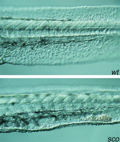
OPTICS: MAGNIFICATION: DATE OF IMAGE: SUBMITTER COMMENTS: Endothelial cells in the ventral fin of mutant (bottom) are not as organized as in wild-type siblings (top). Blood cells accumulate in the empty space in the ventral tail fin of the mutant.
Figure Caption
Fig. for (te382a;te382b)
Original Submitter Comments: Phenotypic class: heart; Visible at: d2; Viability: adult viable
Developmental Stage
Long-pec to Pec-fin
Orientation
| Preparation | Image Form | View | Direction |
| live | still | side view (lateral) | anterior to left |
Figure Data
Acknowledgments
This image is the copyrighted work of the attributed author or publisher, and
ZFIN has permission only to display this image to its users.
Additional permissions should be obtained from the applicable author or publisher of the image.

