
|
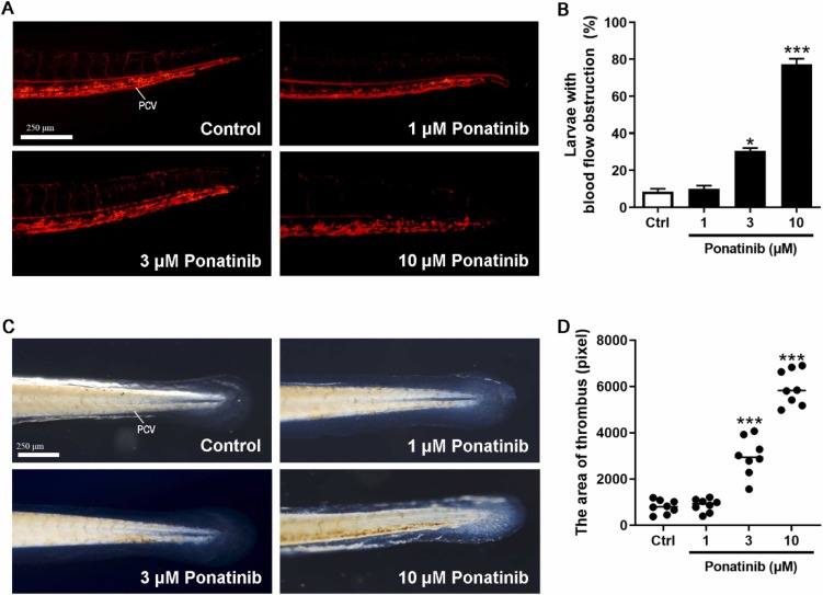
Fig. 3 Thrombosis in Ponatinib-treated zebrafish larvae. (A) 1 dpf Tg (gata1: dsRed) transgenic zebrafish larvae were treated with 0.1 % DMSO or indicated concentrations of Ponatinib for 3 days. The represented images of Ponatinib-induced blood flow stop in the tail of 4dp zebrafish larvae. PCV: Posterior caudal vein. (B) The incidences of blood flow stop in 15 fish/group batches. Data are represented as mean � SEM. *P < 0.05 and ***P < 0.005 vs. control group. (C) The rate of the thrombus occurring in the zebrafish larvae was recorded. Data are presented as means � SD (n = 3). *P < 0.05 and ***P < 0.005 vs. control group. (D) The represented images of Ponatinib-induced thrombus formation (the accumulation of erythrocytes) in 4 dpf zebrafish larvae detected by the o-dianisidine staining. (E) The areas of thrombus of 4 dpf zebrafish were measured by image J (n=8). Data are represented as means � SEM. ***P < 0.005 vs. control group.