
|
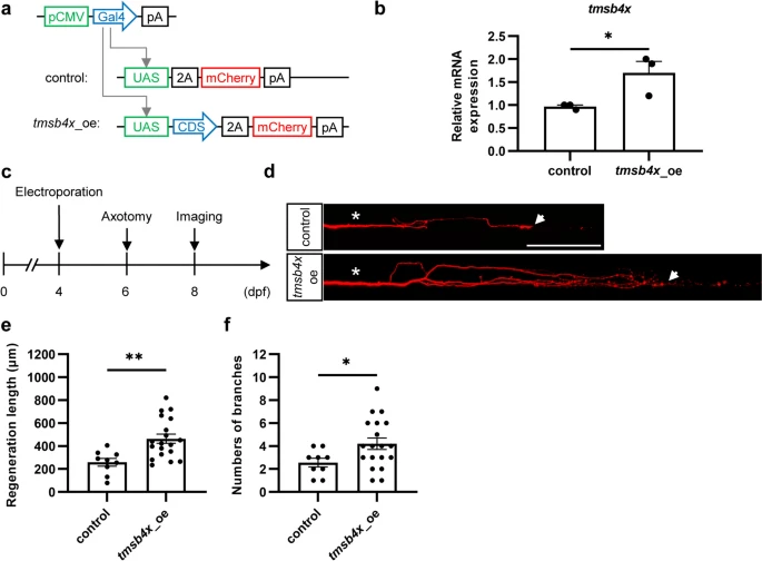
Fig. 3 In vivo single-cell overexpression of tmsb4x promotes axon regeneration in Mauthner cells. a Schematic representation of the overexpression plasmids. b Efficiency of the tmsb4x overexpression plasmid. The data were assessed by an unpaired, two-tailed t test. p = 0.045. c Experimental workflow diagram. d Representative images of axon regeneration after tmsb4x overexpression. Asterisks indicate the sites of injury, and arrows indicate the regenerated axon terminals. Scale bar, 100 ?m. e and f Statistical graphs of the length (e) and branch number (f) of regenerated axons overexpressing tmsb4x (length: control, 258.8 � 34.4 ?m; tmsb4x_oe, 464.1 � 40.0 ?m; branch number: control, 2.6 � 0.4; tmsb4x_oe, 4.2 � 0.5). The results were assessed by an unpaired, two-tailed t test (length, p = 0.0031; branch number, p = 0.042)