
|
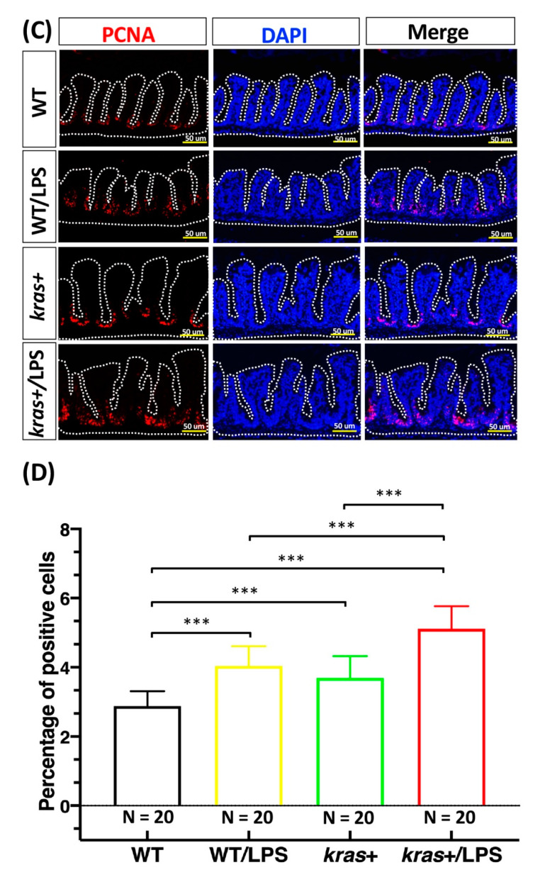
Figure 4
Expression of krasV12 with LPS treatment enhanced the increase in cell apoptosis and cell proliferation in the intestinal epithelium. (A,C) Immunofluorescence staining (red) was carried out in intestinal paraffin sections of WT (N = 20), WT/LPS (N = 20), kras+ (N = 20), and kras+/LPS (N = 20) zebrafish. (B,D) Immunofluorescence staining of caspase-3 showing (1) apoptosis and (2) PCNA as a marker for cell proliferation as well as (3) quantification of the number and percentage of positive cells. Differences among the variables were assessed using Student?s t-tests. Statistical significance: * p < 0.05, *** p < 0.001. Scale bar: 50 ?m.