
|
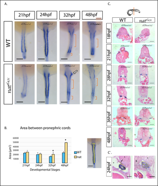
Fig. 3 Renal cells determination occurs normally in nattl43c mutants. A. The pattern of expression from 21 to 48 hpf of Myl7 / ATP?1a.1 gene in situ hybridization. There is no major difference between the expression of WT and mutant embryos. At 32 and 48 hpf, the expression signal is localized at the anterior and medial region of the renal field (brackets), indicative of tissue segmentation. The arrow shows the presumptive Proximal Convoluted Tube (PCT). B. As indicated. n = 8, measurements were done on two independent hybridization experiments. Results presented as mean � s.e.m. p<0.05. Dorsal view, anterior up. Scale bar: 100 ?m C. H&E histology slides on ATP?1a.1 hybridized embryos. Although in most cases the pronephros was absent in mutants, they have renal cells determination as can be evidenced by the ATP?1a.1 expression signal. Dorsal up. Scale bars: 15 ?m. n = 4. Diagram shows the level examined. C`. H&E histology slides on ATP?1a.1 hybridized embryos at 24 hpf show that cells are determined to renal fate but show epithelial abnormalities. WT embryos present normal organized pronephric cell clusters with a lumen and normal anatomical characteristics like Dorsal Aorta (v) and few melanocytic neural crest cells (arrow). nattl43c mutants show disorganized pronephric cells that fail to form lumen and a lower expression signal intensity. There are no melanocytic neural crest cells and there is blood cell accumulation. s (somite), v (vasculature), b (blood cells) Left side, Dorsal up. Scale bars C`: 4 ?m.