
|
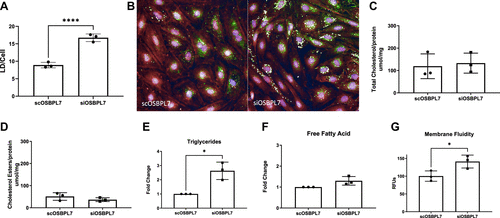
Fig. 3 OSBPL7 deficiency in podocytes alters lipid homeostasis. A: LD quantification per cell in siOSBPL7 podocytes versus control. B: representative images of LD in scOSBPL7 and siOSBPL7 cells stained for LD (green), cytoskeleton (red), and nucleus (blue). Total cholesterol (C) and cholesterol ester levels (D) were not changed between siOSBPL7 and scOSBPL7 podocytes. Triglycerides were increased in siOSBPL7 podocytes (E), while free fatty acids (F) were not changed as indicated by the fold change from siOSBPL7 compared with scOSBPL7 levels. G: membrane fluidity was increased in siOSBPL7 podocytes compared with scOSBPL7. n = 3. *P < 0.05 and ****P < 0.001. LD, lipid droplet; OSBPL7, oxysterol-binding protein-like 7.