
|
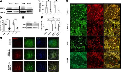
Fig. 1 Decreased OSBPL7 expression in mouse models of experimental CKD is associated with podocyte injury. A and B: representative Western blot images (A) and quantification showing OSBPL7 levels in the kidney cortex of Col4a3?/? and db/db mice compared with Col4a3+/+ (n = 3, *P < 0.05) and db/+ (n = 4, **P < 0.001) littermates (B). C: immunofluorescence staining showcasing OSBPL7 (green) and WT1 (red) in glomeruli, with the merged image highlighting their colocalization, indicating OSBPL7's association with podocytes. D: quantification of OSBPL7 and WT1 colocalization, presented as the ratio of OSBPL7/WT1 double-positive signals to total WT1-positive cells, showing the significant decrease in OSBPL7 in the glomeruli of CKD models compared with controls (n = 10, ****P < 0.0001). E: stable OSBPL7-deficient (SiOSBPL7) podocytes were generated using siRNA. F: apoptosis levels increased in OSBPL7-deficient podocytes and returned to control levels with transfection of full-length OSBPL7 (OSBPL7FL) but not with transfection of OSBPL7 plasmid containing a deletion of the FFAT domain (OSBPL7FFAT?). n = 3 technical replicates. *P < 0.05 and **P < 0.01. G: immunofluorescent images of OSBPL7 (red), ER stain (green), and DAPI (blue) in siOSBPL7 cells with and without OSBPL7FL or OSBPL7FFAT? expressing plasmid. CKD, chronic kidney disease; OSBPL7, oxysterol-binding protein-like 7.