
|
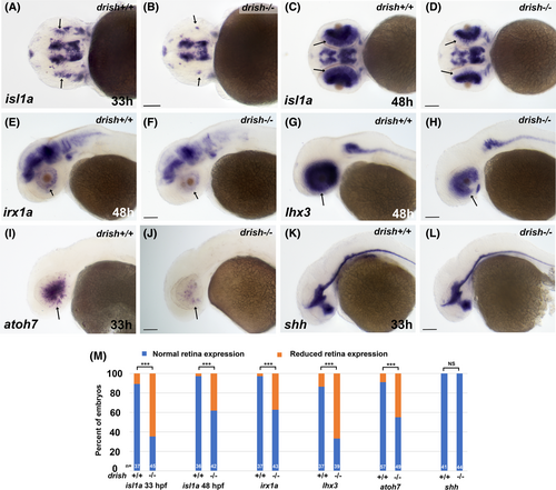
Fig. 7 In situ hybridization analysis of markers associated with RGC differentiation. (A?L) WISH analysis of isl1a, irx1a, lhx3, atoh7, and shh expression in drish+/+ and ?/? embryos at 33 or 48 hpf. Retina specific expression (arrows) of all markers except for shh was reduced in a subset of drish?/? embryos. Scale bar, 50 ?m. (M) Quantification of WISH marker expression. Percentage of wild-type drish+/+ and mutant drish?/? embryos that display retinal expression defects is shown in orange. Data are summary of two independent experiments. The total number of embryos analyzed is shown at the bottom of each bar. ***p < 0.001, Fisher's exact test; NS, not significant.