
|
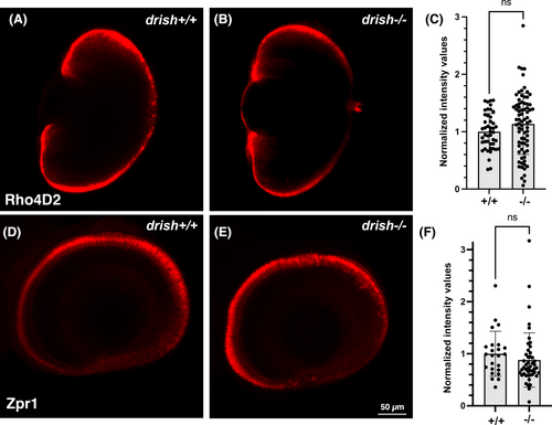
Fig. 6 Immunostaining for photoreceptor marker expression. (A, B) Immunostaining using anti-Rhodopsin antibody Rho4D2 does not show significant changes in drish?/? compared to wild-type larvae at 5 dpf. Ventral view, imaged by confocal microscopy. (C) Quantification of staining intensity in drish?/? and +/+ larvae. ns, not significant, Student's t-test. (D, E) Immunostaining using cone-specific Zpr1 antibody does not show significant changes in drish?/? larvae compared to wild-type at 5 dpf. Lateral view of the eye, imaged by confocal microscopy. Selected slices were combined using maximal-intensity projection. (F) Quantification of staining intensity in drish?/? and +/+ larvae. ns, not significant, Student's t test.