
|
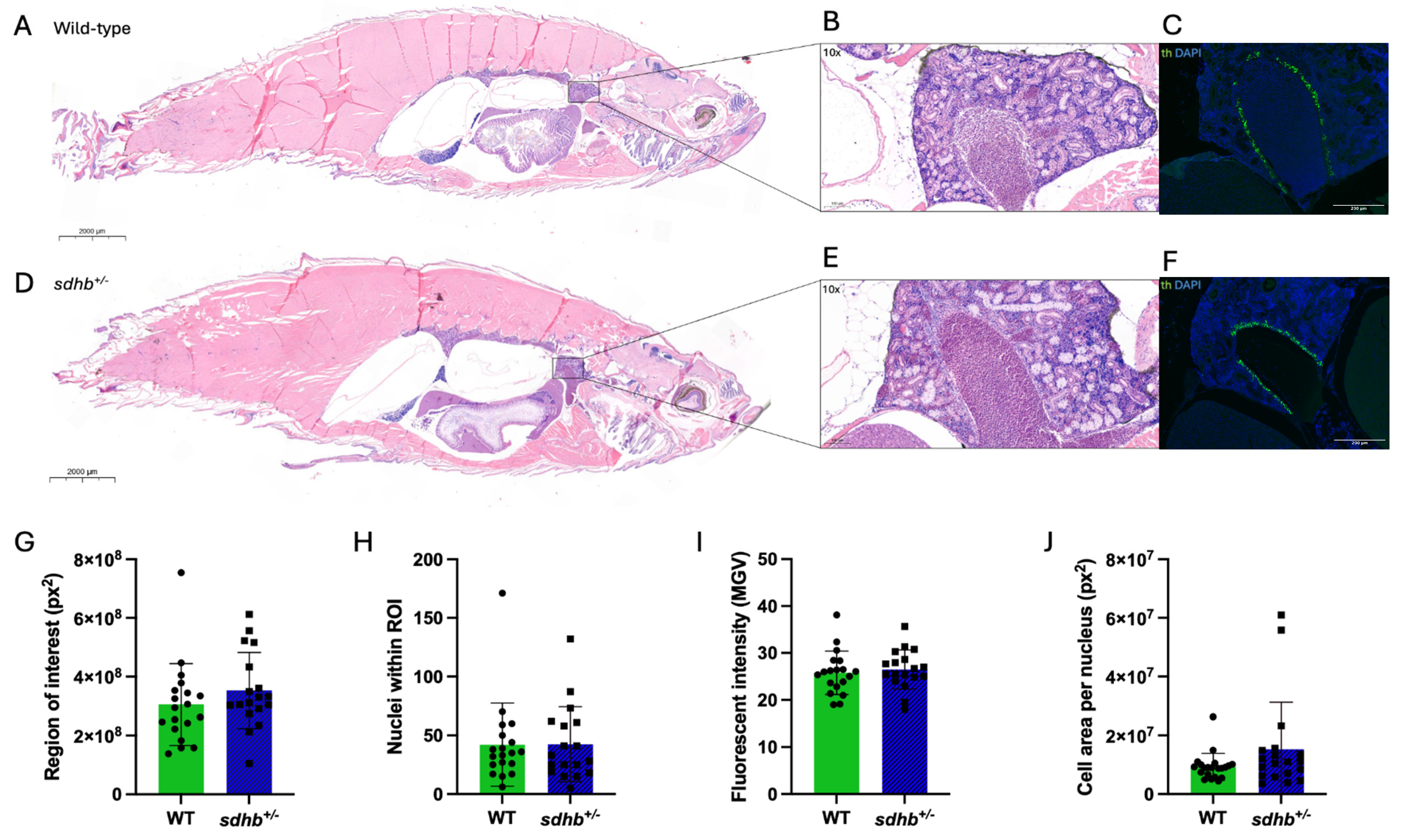
Fig. 3 General histology, kidney morphology, and chromaffin cell morphology of adult heterozygous sdhb mutant and WT zebrafish. (A,D) General histology of adult heterozygous sdhb mutant (n = 34) and WT (n = 35) zebrafish (Representative images; H&E staining, 0.8� magnification). (B,E) Head kidney morphology (H&E staining, 10� magnification). (C,F) Immunohistochemical staining of tyrosine hydroxylase in the head kidney of heterozygous sdhb mutant (n = 10) and WT (n = 9) zebrafish (10� magnification). (G?J) Quantification of tyrosine hydroxylase staining (ImageJ2). Student?s t test was used to evaluate statistical significance. No significant differences were found in any of the analyses.