
|
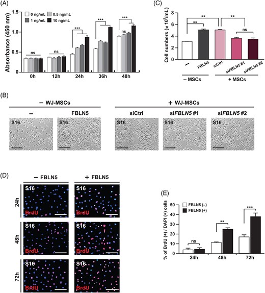
Fig. 2 FBLN5 promotes the proliferation of Schwann cells. A, Proliferating cells in the S16 cells treated with FBLN5 at the indicated concentrations were counted by CCK-8 assay at the indicated incubation times. Statistical significance was determined using 2-way ANOVA followed by Bonferroni's post hoc test (***P < .001, ns: non-significant). B, Images of S16 cells after 24 hours of cultivation with nothing, recombinant FBLN5, or with siRNAs-transfected WJ-MSCs. Scale bars, 400 ?m. C, Quantification of total S16 cell numbers counted for each indicated condition. Statistical significance was determined using one-way ANOVA followed by Tukey's post hoc test (**P < 0.005, ns: non-significant). D, Images of S16 cells with or without recombinant FBLN5, comparing proliferating cells by BrdU assay. Cellular nuclei were stained with DAPI (blue). Scale bars, 400 ?m. E, Quantification of proliferative cells in (D) from each field of view at the indicated time points. Statistical significance was determined using 2-way ANOVA followed by Bonferroni's post hoc test (**P < .005, ***P < .001, ns: non-significant). The data are shown as the mean � SD of three independent experiments per condition