
|
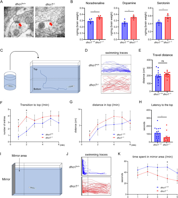
Fig. 4 Synaptic defects and behavioral abnormalities in dhcr7?/? zebrafish (A) Transmission electron micrographs of the telencephalon from 2-mpf dhcr7+/+ and dhcr7?/?zebrafish. (B) Monoamine levels in the brains from 2-mpf dhcr7+/+ and dhcr7?/? zebrafish (n = 4 each). (C) Schematic illustration of the novel tank diving test. (D) Representative swimming traces of 2-mpf dhcr7+/+ and dhcr7?/? zebrafish for the first 2 min of the novel tank diving test. (E) Total swimming distance of 2-mpf dhcr7+/+ (n = 13) and dhcr7?/? (n = 14) zebrafish in 6 min. (F) The number of entries per min to the top half of the tank for 2-mpf dhcr7+/+ (n = 13) and dhcr7?/? (n = 14) zebrafish. (G) Swimming distance per min in the upper half of the tank for 2-mpf dhcr7+/+ (n = 13) and dhcr7?/? (n = 14) zebrafish. (H) Latency entering the tank's top half for 2-mpf dhcr7+/+ (n = 13) and dhcr7?/? (n = 14) zebrafish. (I) Schematic illustration of the mirror biting test. (J) Representative swimming traces of 2-mpf dhcr7+/+ and dhcr7?/? zebrafish for a 5 min mirror-biting test.(K) Time spent per min in the mirror area for 2-mpf dhcr7+/+ (n = 10) and dhcr7?/? (n = 9) zebrafish. All values are presented as the mean � SEM. *p < 0.05, **p < 0.01. n.s: not significant.