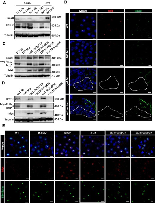
Fig. 3 Protein stability of Rcl1 is dependent on Bms1l protein level in zebrafish. (A) Western blot analysis of Bms1l and Rcl1 protein levels in zebrafish at 5 dpf. Tubulin: loading control. 163 sib, bms1lsq163/+ and WT siblings; 163 MU, bms1lsq163/sq163 mutant; zju1 sib, bms1lzju1/+ or rcl1zju1/+ and the corresponding WT siblings; zju1 MU, bms1lzju1/zju1 or rcl1zju1/zju1 mutant. (B) Immunofluorescence staining of Rcl1 and Bms1l in WT, bms1lsq163/sq163 (163 MU), bms1lzju1/zju1(zju1 MU), and rcl1zju1/zju1 mutant at 5 dpf. Blue signal represents nucleus staining by DAPI. Dotted box indicates liver region. Scale bar, 5 μm. (C and D) Western blot analysis of Bms1l, Rcl1 and Rcl1-Myc protein levels in bms1lsq163/sq163 mutant and siblings (C) or bms1lzju1/zju1 mutant and siblings (D) with or without the background of TgR1# or TgR3# at 5 dpf. (E) Immunofluorescence staining of Myc and the nucleolar marker fibrillarin to demonstrate the localization of Rcl1-Myc in hepatocytes of WT, bms1lsq163/sq163mutant, TgR1#, TgR3#, bms1lsq163/sq163/TgR1#, and bms1lsq163/sq163/TgR3# at 5 dpf. Blue signal represents nucleus staining by DAPI. Scale bar, 5 μm.
Image
Figure Caption
Acknowledgments
This image is the copyrighted work of the attributed author or publisher, and
ZFIN has permission only to display this image to its users.
Additional permissions should be obtained from the applicable author or publisher of the image.
Full text @ J. Mol. Cell Biol.

